Copyright
©The Author(s) 2003.
World J Gastroenterol. Jun 15, 2003; 9(6): 1337-1341
Published online Jun 15, 2003. doi: 10.3748/wjg.v9.i6.1337
Published online Jun 15, 2003. doi: 10.3748/wjg.v9.i6.1337
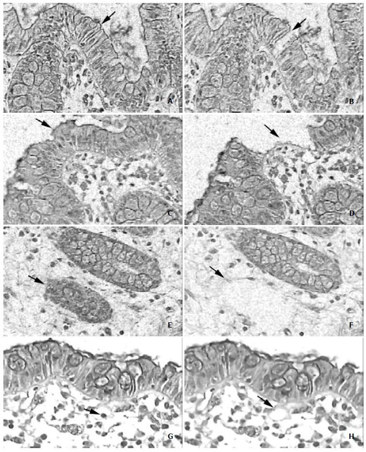
Figure 1 Examples of laser microdissection in colon tissues (hematoxylin and eosin, magnification × 200).
Single cells were picked by combination laser-manipulated microdissection (LMM) and laser pressure catapulting (LPC). A, C, E, G: Before Laser microdissection. B, D, F, H: Using LMM, the laser precisely circumcised selected cells, yielding a cut gap between selected and non-selected areas. Then the selected cells were catapulted using the LPC technique. A-F: Typical images before and after LMM and LPC. G, H: Laser microdissection could also be used to cut out the nucleus of a selected cell.
- Citation: Shi X, Kleeff J, Zhu ZW, Schmied B, Tang WH, Zimmermann A, BÜchler MW, Friess H. Gene-expression analysis of single cells-nested polymerase chain reaction after laser microdissection. World J Gastroenterol 2003; 9(6): 1337-1341
- URL: https://www.wjgnet.com/1007-9327/full/v9/i6/1337.htm
- DOI: https://dx.doi.org/10.3748/wjg.v9.i6.1337


