Copyright
©The Author(s) 2003.
World J Gastroenterol. Jun 15, 2003; 9(6): 1312-1317
Published online Jun 15, 2003. doi: 10.3748/wjg.v9.i6.1312
Published online Jun 15, 2003. doi: 10.3748/wjg.v9.i6.1312
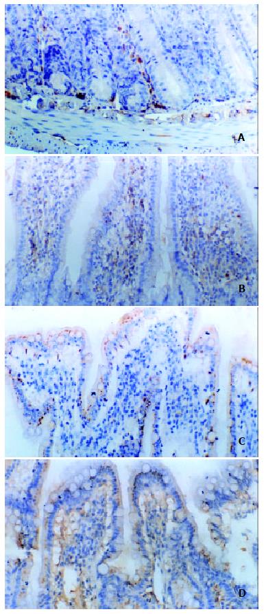
Figure 2 Immunohistochemical staining of phosphorylated p38 MAPK in intestinal biopsies in rats after ischemia/reperfusion injury (SP × 200).
A: Negative control of p38 MAPK staining. There was no positive expression signal in this group; B:Phosphorylated p38 MAPK staining in the saline control group 2 h after reperfusion. Few p38 MAPK positive expression were localized in the cytoplasm and nuclei of villus cells and in the nuclei of crypt cells, mainly in the epithelium and villus cells; C: P38 MAPK staining in the bFGF antibody pre-treated group. The number of positive cells and localization of p38 MAPK positive cells were similar with those in the saline group; D: Phosphorylated p38 staining in the bFGF treated group 2 h after reperfusion. Activated p38 MAPK was localized primarily in the nuclei of crypt cells, few in villus cells. The number of positive cells was more than that in the saline control and bFGF antibody pre-treated groups. In the bFGF treated group, the number of positive expression cells of p38 MAPK as well as its intensity peaked 2 h after reperfusion. ISH × 400
- Citation: Fu XB, Yang YH, Sun TZ, Chen W, Li JY, Sheng ZY. Rapid mitogen-activated protein kinase by basic fibroblast growth factor in rat intestine after ischemia/reperfusion injury. World J Gastroenterol 2003; 9(6): 1312-1317
- URL: https://www.wjgnet.com/1007-9327/full/v9/i6/1312.htm
- DOI: https://dx.doi.org/10.3748/wjg.v9.i6.1312


