Copyright
©The Author(s) 2003.
World J Gastroenterol. Jun 15, 2003; 9(6): 1302-1306
Published online Jun 15, 2003. doi: 10.3748/wjg.v9.i6.1302
Published online Jun 15, 2003. doi: 10.3748/wjg.v9.i6.1302
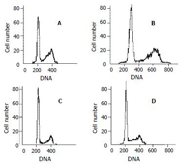
Figure 4 Cell cycle analysis.
Representative flow cytometry data from QBC939 cells after 48 h in the presence of various concentration of celecoxib: 0 μmol/L (A), 10 μmol/L (B), 20 μmol/L (C) and 40 μmol/L (D). The percentage of QBC939 cells in G0-G1 phase after treatment with 40 μmol/L (74.66 ± 6.21) and 20 μmol/L (68.63 ± 4.36) of celecoxib increased significantly compared with control cells (t test, P < 0.01).
-
Citation: Wu GS, Zou SQ, Liu ZR, Tang ZH, Wang JH. Celecoxib inhibits proliferation and induces apoptosis
via prostaglandin E2 pathway in human cholangiocarcinoma cell lines. World J Gastroenterol 2003; 9(6): 1302-1306 - URL: https://www.wjgnet.com/1007-9327/full/v9/i6/1302.htm
- DOI: https://dx.doi.org/10.3748/wjg.v9.i6.1302


