Copyright
©The Author(s) 2003.
World J Gastroenterol. Jun 15, 2003; 9(6): 1191-1195
Published online Jun 15, 2003. doi: 10.3748/wjg.v9.i6.1191
Published online Jun 15, 2003. doi: 10.3748/wjg.v9.i6.1191
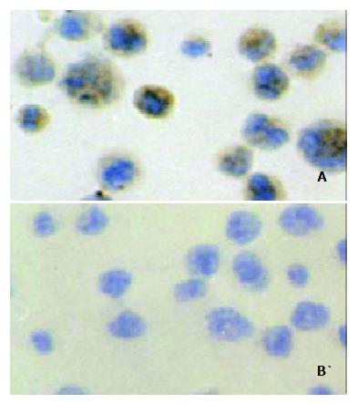
Figure 5 Immunohistochemical staining of the Ehrlich ascites carcinoma cells (EAC): Positive signal was seen in the cytoplasm and membrane of the EAC cells (A).
When stained with a negative control monoclonal antibody (anti-E-tag antibody), the EAC cells showed no positive staining (B).
-
Citation: Guo CC, Ding J, Pan BR, Yu ZC, Han QL, Meng FP, Liu N, Fan DM. Development of an oral DNA vaccine against MG7-Ag of gastric cancer using attenuated
salmonella typhimurium as carrier. World J Gastroenterol 2003; 9(6): 1191-1195 - URL: https://www.wjgnet.com/1007-9327/full/v9/i6/1191.htm
- DOI: https://dx.doi.org/10.3748/wjg.v9.i6.1191


