Copyright
©The Author(s) 2003.
World J Gastroenterol. May 15, 2003; 9(5): 978-983
Published online May 15, 2003. doi: 10.3748/wjg.v9.i5.978
Published online May 15, 2003. doi: 10.3748/wjg.v9.i5.978
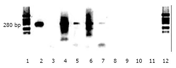
Figure 4 Detection of HBV cccDNA in liver.
Low molecular weight DNA was extracted from liver, digested with Mung Bean nuclease or amplified by PCR without nuclease digestion as described in Materials and Methods. Lane 1, molecular weight markers; lane 2, 5 μg DNA from HepG2 2.2.15 cell media (containing partially double stranded viral DNA); lane 3, 25 μg DNA from HepG2 2.2.15 cell media plus 0.1 U Mung Bean nuclease pretreatment; lane 4, 5 μg DNA from HepG2 2. 2.15 cell layer; lane 5, 5 μg DNA from HepG2 2.2.15 cell layer plus 0.1 U nuclease pretreatment; lane 6, 5 μg liver DNA from a tolerized, transplanted and HBV inoculated rat; lane 7, 5 μg liver DNA from a tolerized, transplanted and HBV inoculated rat; plus 0.1 U nuclease; lane 8, 5 μg liver DNA from a non-transplanted rat inoculated with HBV; lane 9, 5 μg liver DNA from a non-transplanted rat inoculated with HBV plus 0.1 U nuclease; lane 10, 5 μg DNA from an untreated rat; lane 11, 5 μg DNA from an untreated rat plus 0.1 U nuclease; lanes 1 and 12, molecular size markers.
- Citation: Wu CH, Ouyang EC, Walton C, Promrat K, Forouhar F, Wu GY. Hepatitis B virus infection of transplanted human hepatocytes causes a biochemical and histological hepatitis in immunocompetent rats. World J Gastroenterol 2003; 9(5): 978-983
- URL: https://www.wjgnet.com/1007-9327/full/v9/i5/978.htm
- DOI: https://dx.doi.org/10.3748/wjg.v9.i5.978


