Copyright
©The Author(s) 2003.
World J Gastroenterol. May 15, 2003; 9(5): 1014-1019
Published online May 15, 2003. doi: 10.3748/wjg.v9.i5.1014
Published online May 15, 2003. doi: 10.3748/wjg.v9.i5.1014
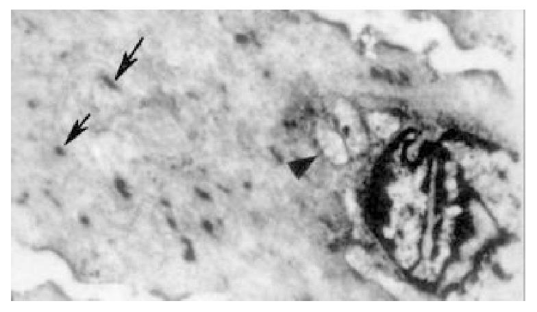
Figure 8 The figures is a transection of cCS portion of bile duct in experimental group.
Under the electronmicroscope the cell membrane twisted like billows, and the myofilament density was not even, shaping like "whirl". The dense body showed derangement and various shapes (↑). The mitochondrions clustered at the end of the nucleus, and vacuolation (▲) was seen.
- Citation: Wei JG, Wang YC, Liang GM, Wang W, Chen BY, Xu JK, Song LJ. The study between the dynamics and the X-ray anatomy and regularizing effect of gallbladder on bile duct sphincter of the dog. World J Gastroenterol 2003; 9(5): 1014-1019
- URL: https://www.wjgnet.com/1007-9327/full/v9/i5/1014.htm
- DOI: https://dx.doi.org/10.3748/wjg.v9.i5.1014


