Copyright
©The Author(s) 2003.
World J Gastroenterol. Apr 15, 2003; 9(4): 645-649
Published online Apr 15, 2003. doi: 10.3748/wjg.v9.i4.645
Published online Apr 15, 2003. doi: 10.3748/wjg.v9.i4.645
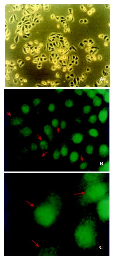
Figure 3 The localization of annexin I in ESCC cell line.
EC0156 cells were grown on glass coverslips at 80% confluence, de-tected with the anti-annexin I monoclonal antibody and visu-alized by FITC-conjugated secondary antibody under the fluo-rescence microscope. EC0156 cells were cultured in DMEM medium and photomicrographied under a phase-contrast mi-croscopy (a 100 ×). Annexin I was observed by fluorescence microscope (b 400 × and c 1000 ×). The red arrows showed that annexin I protein was also located on nuclear membranes in EC0156 cells.
- Citation: Liu Y, Wang HX, Lu N, Mao YS, Liu F, Wang Y, Zhang HR, Wang K, Wu M, Zhao XH. Translocation of annexin I from cellular membrane to the nuclear membrane in human esophageal squamous cell carcinoma. World J Gastroenterol 2003; 9(4): 645-649
- URL: https://www.wjgnet.com/1007-9327/full/v9/i4/645.htm
- DOI: https://dx.doi.org/10.3748/wjg.v9.i4.645


