Copyright
©The Author(s) 2003.
World J Gastroenterol. Apr 15, 2003; 9(4): 645-649
Published online Apr 15, 2003. doi: 10.3748/wjg.v9.i4.645
Published online Apr 15, 2003. doi: 10.3748/wjg.v9.i4.645
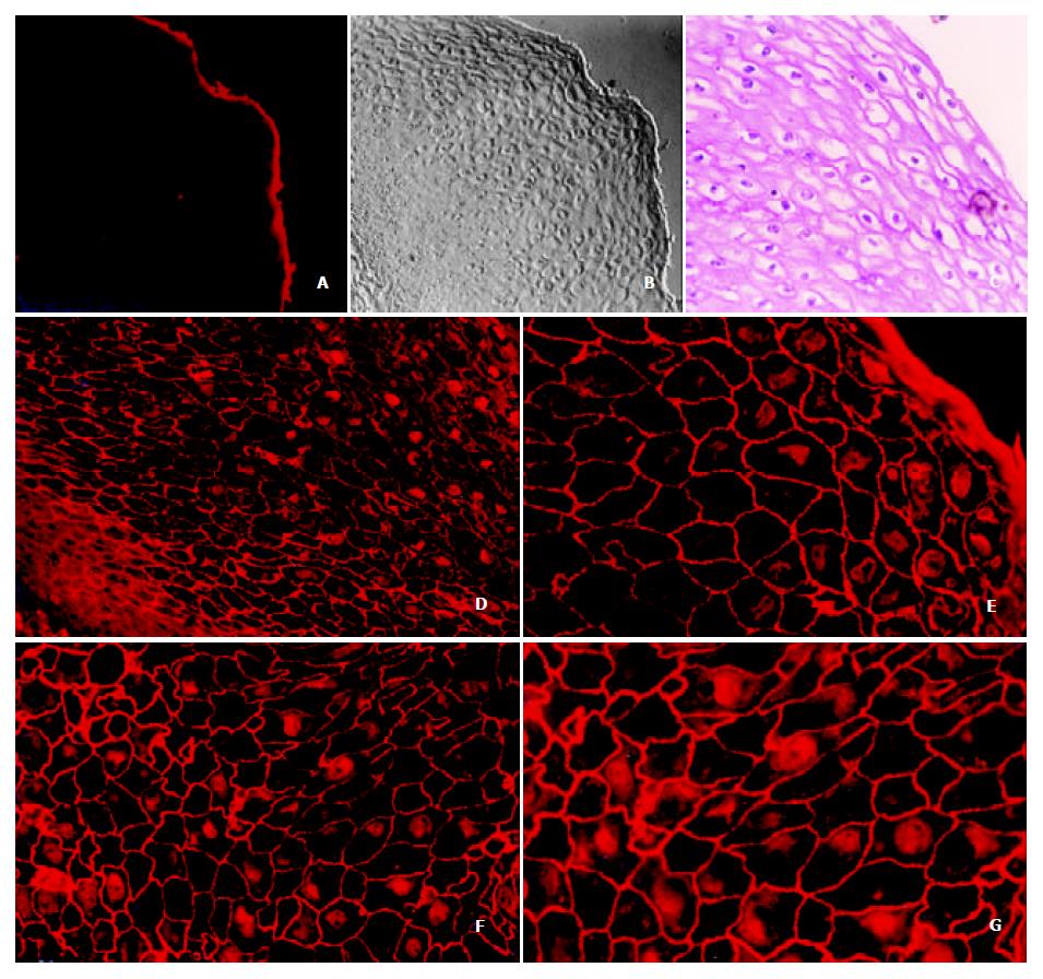
Figure 1 The localization of annexin I in normal esophageal epithelium.
The normal esophageal epithelia on the paraffin-embed-ded tissues were detected with anti-annexin I monoclonal antibody and imaged with TRITC-conjugated goat anti-mouse antibody, and then observed under a fluorescence microscope (a, d, e, f and g). PBS was used as a negative control (a 50 ×), the tissue section was viewed under a Nomarski interference-contrast microscopy (b 50 ×) and the additional H & E staining was also performed (c 200 ×). The localization of annexin I protein in normal esophageal epithelium was shown from d to g (d 200 ×; e 400 ×; f 400 × and g 400 ×). The orientation of the bottom left corner was the basal membrane of the esophageal mucosa.
- Citation: Liu Y, Wang HX, Lu N, Mao YS, Liu F, Wang Y, Zhang HR, Wang K, Wu M, Zhao XH. Translocation of annexin I from cellular membrane to the nuclear membrane in human esophageal squamous cell carcinoma. World J Gastroenterol 2003; 9(4): 645-649
- URL: https://www.wjgnet.com/1007-9327/full/v9/i4/645.htm
- DOI: https://dx.doi.org/10.3748/wjg.v9.i4.645


