Copyright
©The Author(s) 2003.
World J Gastroenterol. Mar 15, 2003; 9(3): 627-630
Published online Mar 15, 2003. doi: 10.3748/wjg.v9.i3.627
Published online Mar 15, 2003. doi: 10.3748/wjg.v9.i3.627
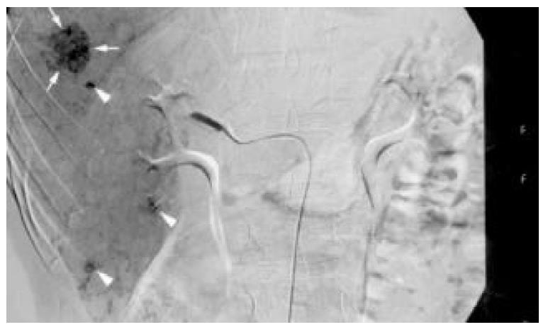
Figure 3 On angiography, there was an oval hypervascular lesion (black arrows) and multiple scattered tiny hypervascular spots in the right lobe of the liver (white arrows).
- Citation: Hsu CY, Chu CH, Lin SC, Yang FS, Yang TL, Chang KM. Concomitant hepatocellular adenoma and adenomatous hyperplasia in a patient without cirrhosis. World J Gastroenterol 2003; 9(3): 627-630
- URL: https://www.wjgnet.com/1007-9327/full/v9/i3/627.htm
- DOI: https://dx.doi.org/10.3748/wjg.v9.i3.627


