Copyright
©The Author(s) 2003.
World J Gastroenterol. Feb 15, 2003; 9(2): 258-261
Published online Feb 15, 2003. doi: 10.3748/wjg.v9.i2.258
Published online Feb 15, 2003. doi: 10.3748/wjg.v9.i2.258
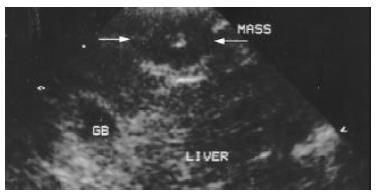
Figure 1 Image of VX2 tumor lesion under conventional B mode US.
Arrow indicates the VX2 tumor lesion at the anterior part of the right lobe. It is oval and hypoechoic with a small hyperechoic scar at the center of the lesion.
- Citation: Du WH, Yang WX, Wang X, Xiong XQ, Zhou Y, Li T. Vascularity of hepatic VX2 tumors of rabbits: Assessment with conventional power Doppler US and contrast enhanced harmonic power Doppler US. World J Gastroenterol 2003; 9(2): 258-261
- URL: https://www.wjgnet.com/1007-9327/full/v9/i2/258.htm
- DOI: https://dx.doi.org/10.3748/wjg.v9.i2.258


