Copyright
©The Author(s) 2003.
World J Gastroenterol. Dec 15, 2003; 9(12): 2828-2831
Published online Dec 15, 2003. doi: 10.3748/wjg.v9.i12.2828
Published online Dec 15, 2003. doi: 10.3748/wjg.v9.i12.2828
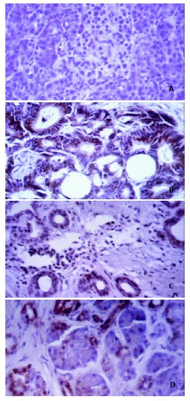
Figure 1 Immunohistochemical staining of pancreatic tissues using antiserum against human Caspase-1.
A: Normal pancreas showed only occasional slight staining. B: Pancreatic cancer cells showed a clear cytoplasmatic immunoreactivity in 71%. C: Hyperplastic ducts in chronic pancreatitis tissues also showed cytoplasmatic staining in 87% of the tissues, whereas in atrophic acinar cells (D) a predominantly nuclear staining could be observed in 89%.
- Citation: Yang YM, Ramadani M, Huang YT. Overexpression of Caspase-1 in adenocarcinoma of pancreas and chronic pancreatitis. World J Gastroenterol 2003; 9(12): 2828-2831
- URL: https://www.wjgnet.com/1007-9327/full/v9/i12/2828.htm
- DOI: https://dx.doi.org/10.3748/wjg.v9.i12.2828


