Copyright
©The Author(s) 2003.
World J Gastroenterol. Dec 15, 2003; 9(12): 2751-2758
Published online Dec 15, 2003. doi: 10.3748/wjg.v9.i12.2751
Published online Dec 15, 2003. doi: 10.3748/wjg.v9.i12.2751
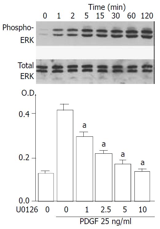
Figure 7 PDGF-BB induced proliferation of SIPS cells through ERK pathway.
(A) SIPS cells were treated with PDGF-BB (at 25 ng/ml) for the indicated time. Total cell lysates (approximately 100 μg) were prepared, and the level of activated, phosphorylated ERK was determined by Western blotting. The level of total ERK was also determined. (B) SIPS cells were treated with PDGF-BB (at 25 ng/ml) in the presence of U0126 at the indicated concentrations (at μM). After 24-hour-incubation, cells were labeled with BrdU for 3 hours. Cells were fixed, and incubated with peroxidase-conjugated anti-BrdU antibody. Then the peroxidase substrate 3,3’,5,5’-tetramethylbenzidine was added, and BrdU incorporation was quantitated by differences in absorbance at O.D.370-O.D.492. Data are shown as mean ± SD (n = 6). aP < 0.01 versus PDGF only. O.D.: optical density.
- Citation: Masamune A, Satoh M, Kikuta K, Suzuki N, Shimosegawa T. Establishment and characterization of a rat pancreatic stellate cell line by spontaneous immortalization. World J Gastroenterol 2003; 9(12): 2751-2758
- URL: https://www.wjgnet.com/1007-9327/full/v9/i12/2751.htm
- DOI: https://dx.doi.org/10.3748/wjg.v9.i12.2751


