Copyright
©The Author(s) 2003.
World J Gastroenterol. Dec 15, 2003; 9(12): 2751-2758
Published online Dec 15, 2003. doi: 10.3748/wjg.v9.i12.2751
Published online Dec 15, 2003. doi: 10.3748/wjg.v9.i12.2751
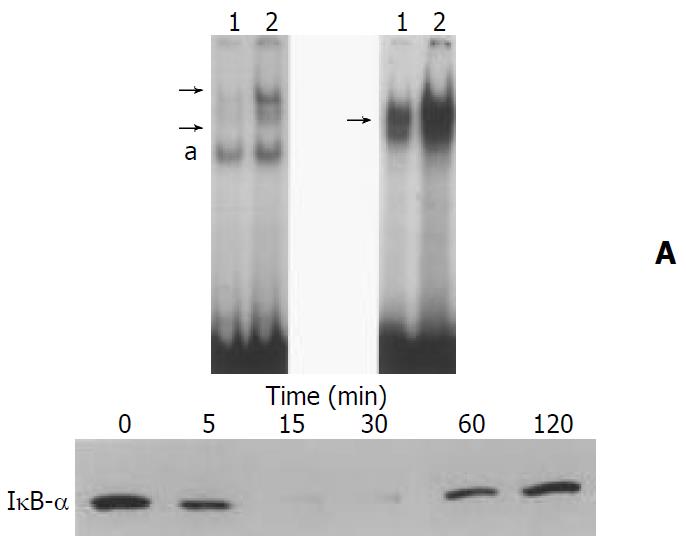
Figure 5 IL-1β activated NF-κB and AP-1 in SIPS cells.
(A, B) SIPS cells were treated with IL-1β (at 2 ng/ml, lane 2) for 1 hour. Nuclear extracts were prepared, and specific binding activities of NF-κB (panel A) and AP-1 (panel B) were assessed by electro-phoretic mobility shift assay. Arrows denote specific inducible complexes competitive with cold double-stranded oligonucle-otide probes. Lane 1: control (medium only). a: non-specific band. (C) SIPS cells were treated with IL-1β for indicated times. Total cell lysates (approximately 100 μg) were prepared, and the level of IκB-α was determined by Western blotting.
- Citation: Masamune A, Satoh M, Kikuta K, Suzuki N, Shimosegawa T. Establishment and characterization of a rat pancreatic stellate cell line by spontaneous immortalization. World J Gastroenterol 2003; 9(12): 2751-2758
- URL: https://www.wjgnet.com/1007-9327/full/v9/i12/2751.htm
- DOI: https://dx.doi.org/10.3748/wjg.v9.i12.2751


