Copyright
©The Author(s) 2002.
World J Gastroenterol. Dec 15, 2002; 8(6): 1045-1049
Published online Dec 15, 2002. doi: 10.3748/wjg.v8.i6.1045
Published online Dec 15, 2002. doi: 10.3748/wjg.v8.i6.1045
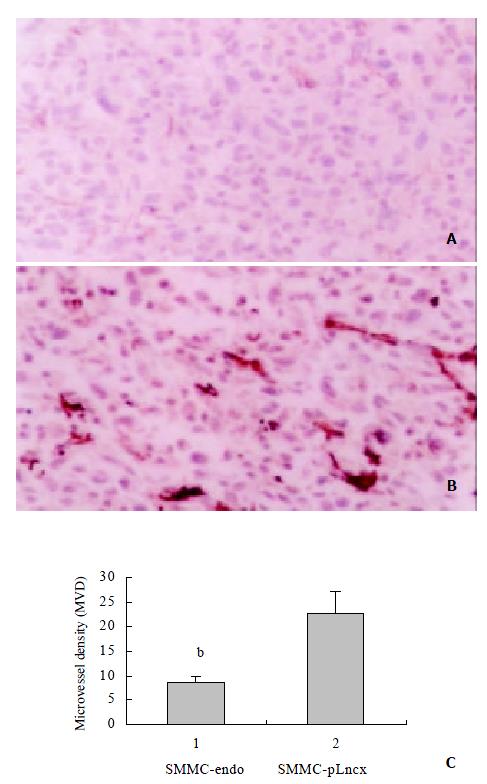
Figure 5 Tumor sections were stained with an antibody reac-tive to CD34.
A: Tumor section from endostatin-transfected g1roup showed only a few positively stained vascular endothelial cells. B: Similar section of the control group showed highly vasuclarized tumor tissue. C: Microvessel density (MVD) was quantified by counting of positively stained endothelial cells from 5 fields in each tumor section. Bars, SD. bP < 0.01, compared with control SMMC-pLncx. × 200.
- Citation: Wang X, Liu FK, Li X, Li JS, Xu GX. Retrovirus-mediated gene transfer of human endostatin inhibits growth of human liver carcinoma cells SMMC7721 in nude mice. World J Gastroenterol 2002; 8(6): 1045-1049
- URL: https://www.wjgnet.com/1007-9327/full/v8/i6/1045.htm
- DOI: https://dx.doi.org/10.3748/wjg.v8.i6.1045


