Copyright
©The Author(s) 2002.
World J Gastroenterol. Aug 15, 2002; 8(4): 638-643
Published online Aug 15, 2002. doi: 10.3748/wjg.v8.i4.638
Published online Aug 15, 2002. doi: 10.3748/wjg.v8.i4.638
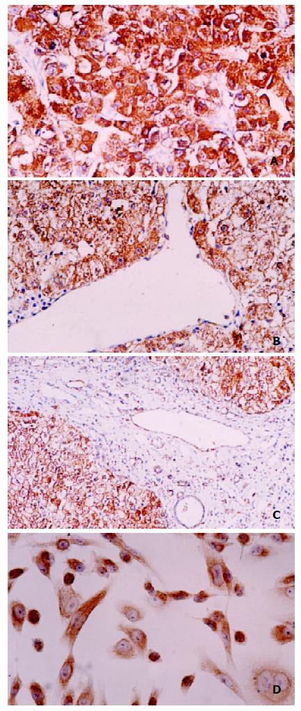
Figure 3 Immunohistochemical staining of P28/Gankyrin in HCC, para-carcinoma liver tissue and SK-Hep1 cell line.
(A) p28/gankyrin was moderately to intensely present in the cytoplasm of most hepatocytes in HCC tissue, and occasionally present in the nucleus of hepatocytes; (B) In contrast, p28/gankyrin was weakly present in the cytoplasm of hepatocytes in para-carcinoma liver tissue; (C) p28/gankyrin protein signals were specifically present in the cytoplasm of hepatocytes, while absent in bile duct cells, blood vein endometrial cells and other interstitial cells; (D) In SK-Hep1 cells, p28/gankyrin was mostly located in cytoplasm and occasionally in the nucleus.
-
Citation: Fu XY, Wang HY, Tan L, Liu SQ, Cao HF, Wu MC. Overexpression of
p 28/gankyrin in human hepatocellular carcinoma and its clinical significance. World J Gastroenterol 2002; 8(4): 638-643 - URL: https://www.wjgnet.com/1007-9327/full/v8/i4/638.htm
- DOI: https://dx.doi.org/10.3748/wjg.v8.i4.638


