Copyright
©The Author(s) 2001.
World J Gastroenterol. Oct 15, 2001; 7(5): 612-621
Published online Oct 15, 2001. doi: 10.3748/wjg.v7.i5.612
Published online Oct 15, 2001. doi: 10.3748/wjg.v7.i5.612
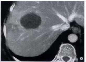
Figure 8 Radiofrequency ablation: the post treatment CT scan shows a 4.
5 cm necrotic RFA lesion at the site of the previous tumour (Figur 7). There is typical marginal enhancement and some perfusional anomalies in the subtended liver.
- Citation: Makin GB, Breen DJ, Monson JR. The impact of new technology on surgery for colorectal cancer. World J Gastroenterol 2001; 7(5): 612-621
- URL: https://www.wjgnet.com/1007-9327/full/v7/i5/612.htm
- DOI: https://dx.doi.org/10.3748/wjg.v7.i5.612


