Copyright
©The Author(s) 2001.
World J Gastroenterol. Jun 15, 2001; 7(3): 422-424
Published online Jun 15, 2001. doi: 10.3748/wjg.v7.i3.422
Published online Jun 15, 2001. doi: 10.3748/wjg.v7.i3.422
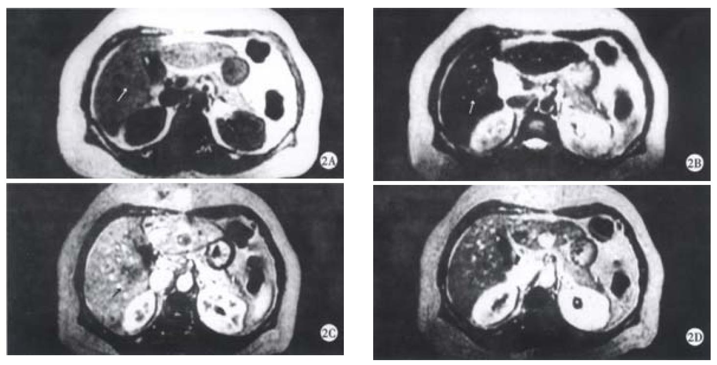
Figure 2 Inflammatory pseudotumor of the liver in the right posterior lobe.
A: SE T1WI showed a hypointense lesion(arrow). B: SE T2WI showed the lesion was slightly and inhomogeneously hyperintense(arrow). C: There is no enhancement in early arterial phase of dynamic contrast MRI, the edge of the lesion was not clear(arrow). D: portal venous phase of dynamic contrast MRI showed the punctual core in the center and peripheral enhancement (arrow).
- Citation: Yan FH, Zhou KR, Jiang YP, Shi WB. Inflammatory pseudotumor of the liver: 13 cases of MRI findings. World J Gastroenterol 2001; 7(3): 422-424
- URL: https://www.wjgnet.com/1007-9327/full/v7/i3/422.htm
- DOI: https://dx.doi.org/10.3748/wjg.v7.i3.422


