Copyright
©The Author(s) 2001.
World J Gastroenterol. Feb 15, 2001; 7(1): 74-79
Published online Feb 15, 2001. doi: 10.3748/wjg.v7.i1.74
Published online Feb 15, 2001. doi: 10.3748/wjg.v7.i1.74
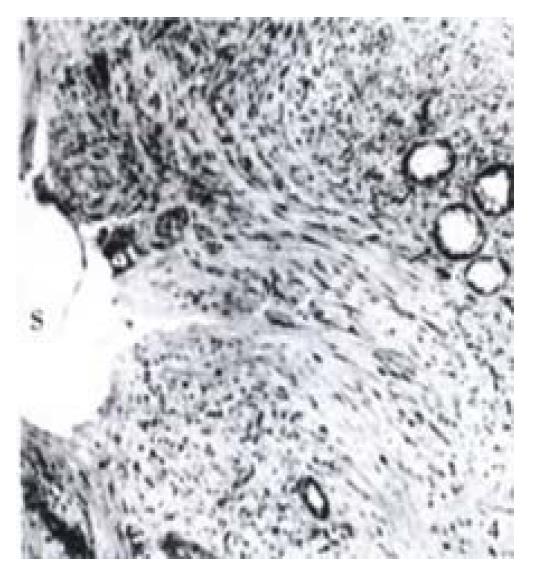
Figure 4 Photomicrography from an occluded TIPS shunt using a Wallstent shows that a massive pseudointimal proliferative tissue under the stent (S), which primarily composed of myofibroblastic cells (M).
Neo-bile duct proliferation within the proliferation is also noted (arrows). Modified Giemsa and basic Fuchsin stain, × 125
- Citation: Teng GJ, Bettmann MA, Hoopes PJ, Yang L. Comparison of a new stent and Wallstent for transjugular intrahepatic portosystemic shunt in a porcine model. World J Gastroenterol 2001; 7(1): 74-79
- URL: https://www.wjgnet.com/1007-9327/full/v7/i1/74.htm
- DOI: https://dx.doi.org/10.3748/wjg.v7.i1.74


