Copyright
©The Author(s) 2000.
World J Gastroenterol. Aug 15, 2000; 6(4): 513-521
Published online Aug 15, 2000. doi: 10.3748/wjg.v6.i4.513
Published online Aug 15, 2000. doi: 10.3748/wjg.v6.i4.513
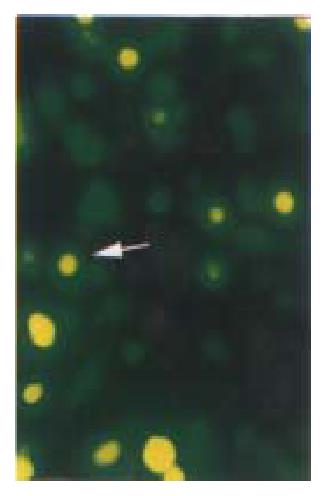
Figure 3 Cell slides under Nikon Eclipse TE200 fluorescence microscope using excitation light at either 488 nm to detect GFP (emission filter 522 nm).
Distribution of GFP p27KIP1 fusion protein expressed in living HCC-9204 cells 8 after 100 μmol/L ZnSO4 treatment, indicated by an arrow. × 200
- Citation: Li J, Yang XK, Yu XX, Ge ML, Wang WL, Zhang J, Hou YD. Overexpression of p27KIP1 induced cell cycle arrest in G1 phase and subsequent apoptosis in HCC-9204 cell line. World J Gastroenterol 2000; 6(4): 513-521
- URL: https://www.wjgnet.com/1007-9327/full/v6/i4/513.htm
- DOI: https://dx.doi.org/10.3748/wjg.v6.i4.513


