Copyright
©The Author(s) 2000.
World J Gastroenterol. Jun 15, 2000; 6(3): 461-464
Published online Jun 15, 2000. doi: 10.3748/wjg.v6.i3.461
Published online Jun 15, 2000. doi: 10.3748/wjg.v6.i3.461
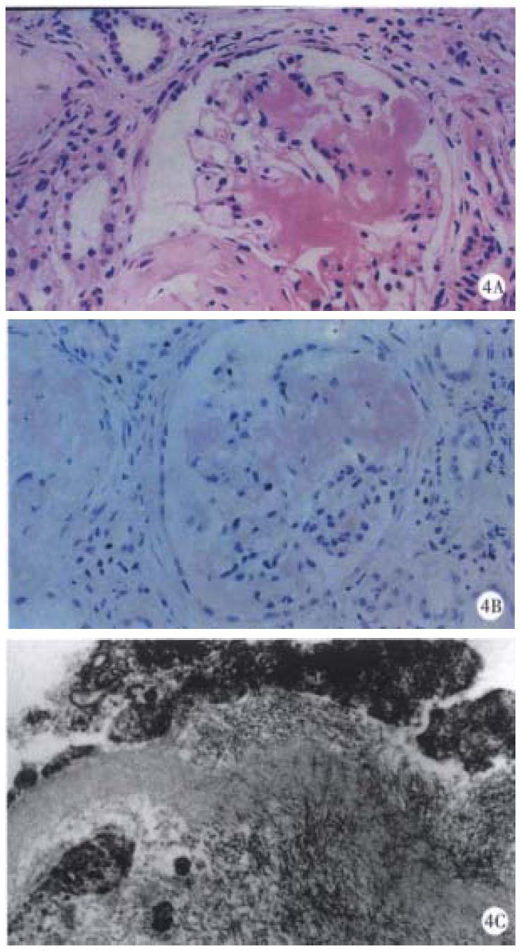
Figure 4 Findings of the renal biopsy.
A: Histological findings (hematocylin and eosin). Amorphous, eosin-stained deposits were seen in the mesangial areas. B: Histological findings (Congo red stain). The deposits were Congo red positive. Congo red stain showed reddish pink deposits that demonstrated apple-green birefringence when examined under polarized light. C: Electron microscopic findings. Fine fibrils (8 to 10 nm in diameter) arranged randomly or in bundles were found in the mesangium.
- Citation: Saitoh O, Kojima K, Teranishi T, Nakagawa K, Kayazawa M, Nanri M, Egashira Y, Hirata I, Katsu KI. Renal amyloidosis as a late complication of Crohn's disease: a case report and review of the literature from Japan. World J Gastroenterol 2000; 6(3): 461-464
- URL: https://www.wjgnet.com/1007-9327/full/v6/i3/461.htm
- DOI: https://dx.doi.org/10.3748/wjg.v6.i3.461


