Copyright
©The Author(s) 2000.
World J Gastroenterol. Apr 15, 2000; 6(2): 210-215
Published online Apr 15, 2000. doi: 10.3748/wjg.v6.i2.210
Published online Apr 15, 2000. doi: 10.3748/wjg.v6.i2.210
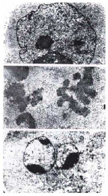
Figure 4 Ultrastructures of SMMC-7721 cells with or without treatment by taxol.
Following 24 h treatment of taxol, cells presented more mitotic Figure (B 7680 ×). For 48 h, the cells appeared with regularly shaped crescents (C 7680 ×). The nuclei of untreated cells were very irregular in shape, with many gulfs and protrusions. (A 5760 ×)
- Citation: Yuan JH, Zhang RP, Zhang RG, Guo LX, Wang XW, Luo D, Xie Y, Xie H. Growth-inhibiting effects of taxol on human liver cancer in vitro and in nude mice. World J Gastroenterol 2000; 6(2): 210-215
- URL: https://www.wjgnet.com/1007-9327/full/v6/i2/210.htm
- DOI: https://dx.doi.org/10.3748/wjg.v6.i2.210


