Copyright
©The Author(s) 2000.
World J Gastroenterol. Feb 15, 2000; 6(1): 102-106
Published online Feb 15, 2000. doi: 10.3748/wjg.v6.i1.102
Published online Feb 15, 2000. doi: 10.3748/wjg.v6.i1.102
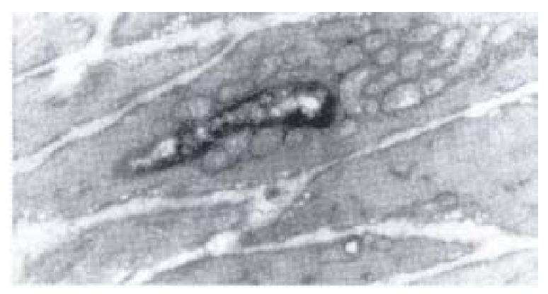
Figure 7 Electron-microscopic scanning of a group I rabbit’s SO smooth muscle.
Image showed the swelling of plasmosome and disap pear of intercristal space, decrease of kink macula densa and twisting of myofil aments.
- Citation: Wei JG, Wang YC, Du F, Yu HJ. Dynamic and ultrastructural study of sphincter of Oddi in early-stage cholelithiasis in rabbits with hypercholesterolemia. World J Gastroenterol 2000; 6(1): 102-106
- URL: https://www.wjgnet.com/1007-9327/full/v6/i1/102.htm
- DOI: https://dx.doi.org/10.3748/wjg.v6.i1.102


