Copyright
©The Author(s) 1999.
World J Gastroenterol. Dec 15, 1999; 5(6): 492-505
Published online Dec 15, 1999. doi: 10.3748/wjg.v5.i6.492
Published online Dec 15, 1999. doi: 10.3748/wjg.v5.i6.492
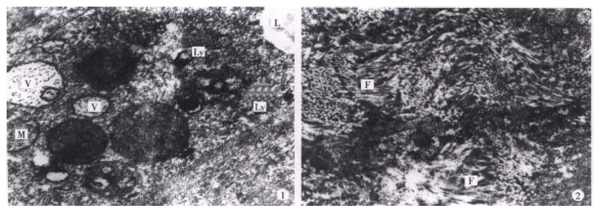
Figure 9 Male, aged 55, hospital no.
202295, clinical diagnosis: left lobe medium sized hepatic carcinoma (5 cm × 6 cm) near t he hepatic hilus, treated by TAE and regimen B, with interval of 2 months, and left hepatic lobectomy was performed 6 months later. The tumor (4 cm × 4 cm) was with hard consistency, and encapsulated fibrotic degeneration . Most of the cancer cells showed necrosis under light microscopy. Some cancer c ells in the cholecystic wall were alive (may be due to the blood supply from cho lecystic artery). The electron microscopy showed: 1, cancer cells degenerated and necrosed; in cytoplasm some vacuolized mitochondria (M), irregular sized vacuol es (V) and lysosome (Ly) and lipid droplet (L) were seen. The rest struct ures were not clear ( × 30000). 2, Prominent fibro-connective tissue hyperplasi a (F) in the tumor tissue ( × 10000) was found. Patient gained body weight and no subjective upset on follow up until 12 mos after operation.
- Citation: Liu L, Jiang Z, Teng GJ, Song JZ, Zhang DS, Guo QM, Fang W, He SC, Guo JH. Clinical and experimental study on regional administration of phosphorus 32 glass microspheres in treating hepatic carcinoma. World J Gastroenterol 1999; 5(6): 492-505
- URL: https://www.wjgnet.com/1007-9327/full/v5/i6/492.htm
- DOI: https://dx.doi.org/10.3748/wjg.v5.i6.492


