Copyright
©The Author(s) 1999.
World J Gastroenterol. Oct 15, 1999; 5(5): 383-387
Published online Oct 15, 1999. doi: 10.3748/wjg.v5.i5.383
Published online Oct 15, 1999. doi: 10.3748/wjg.v5.i5.383
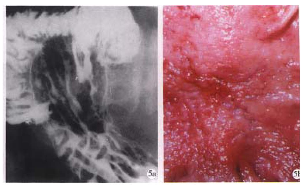
Figure 5 Linear ulcer.
A long linear ulcer paralleled to the lesser curvature (arrowhead) shown on the gastric posterior wall, representing the healing and healed stage of ulcer. B. Macrograph shows that the fine curve linear ulcer is composed of re-epithelialized (healed) and granulating part (healing).
- Citation: Chen JR. Reassessment of barium radiographic examination in diagnosing gastrointestinal diseases. World J Gastroenterol 1999; 5(5): 383-387
- URL: https://www.wjgnet.com/1007-9327/full/v5/i5/383.htm
- DOI: https://dx.doi.org/10.3748/wjg.v5.i5.383


