Copyright
©The Author(s) 1998.
World J Gastroenterol. Jun 15, 1998; 4(3): 214-218
Published online Jun 15, 1998. doi: 10.3748/wjg.v4.i3.214
Published online Jun 15, 1998. doi: 10.3748/wjg.v4.i3.214
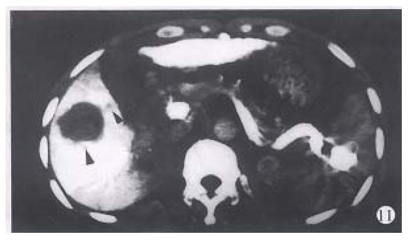
Figure 11 The contrast medium remained in the spleen after CTSP.
The splenic veins show high density. The recurrent cancer in the liver and the child focus in the portal vein (the left posterior arrow) are both shown clearly.
- Citation: Zhang XL, Qiu SJ, Chang RM, Zou CJ. Animal experiments and clinical application of CT during percutaneous splenoportography. World J Gastroenterol 1998; 4(3): 214-218
- URL: https://www.wjgnet.com/1007-9327/full/v4/i3/214.htm
- DOI: https://dx.doi.org/10.3748/wjg.v4.i3.214


