Copyright
©The Author(s) 1998.
World J Gastroenterol. Jun 15, 1998; 4(3): 214-218
Published online Jun 15, 1998. doi: 10.3748/wjg.v4.i3.214
Published online Jun 15, 1998. doi: 10.3748/wjg.v4.i3.214
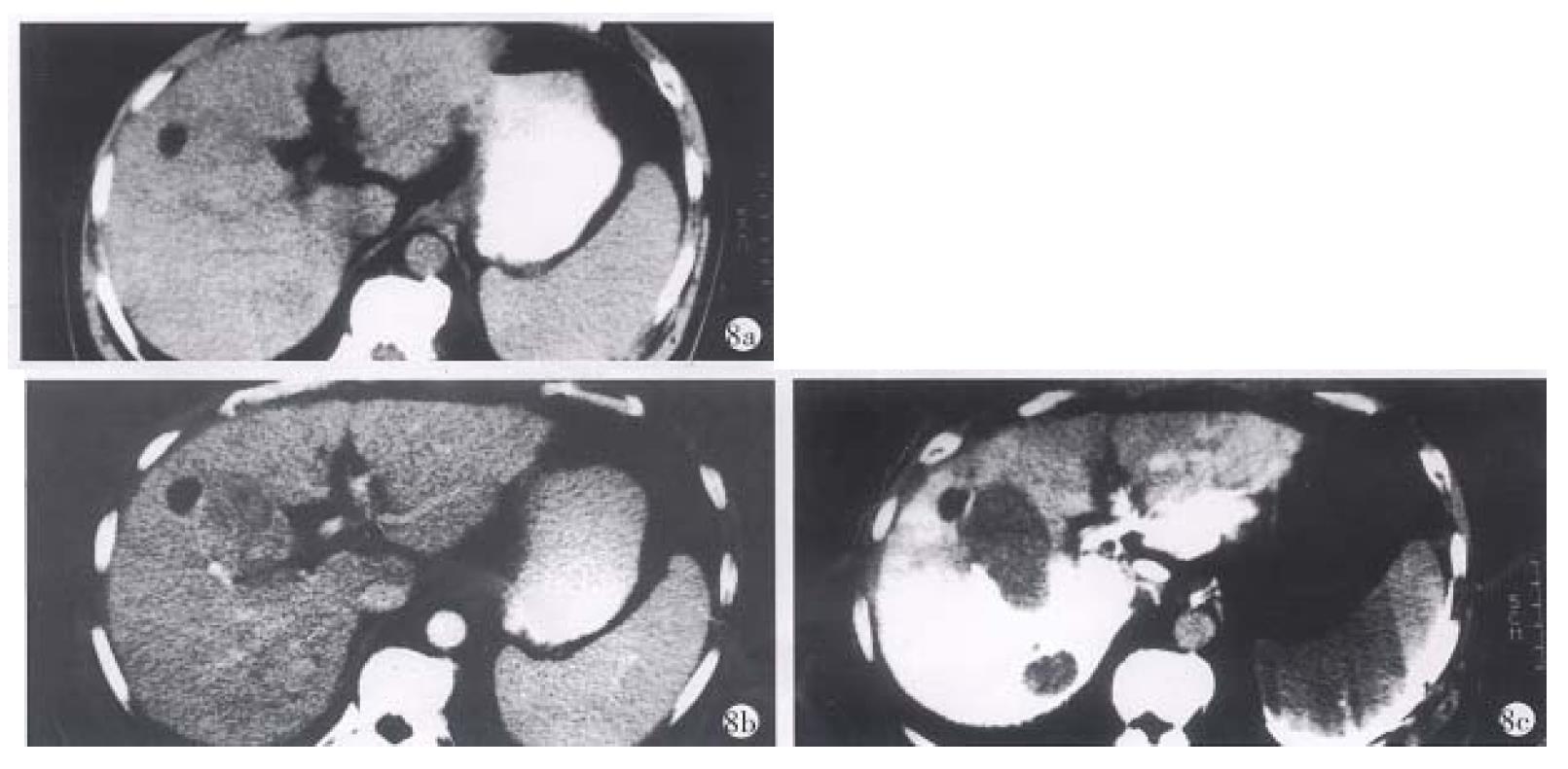
Figure 8 PHC of a 59-year-old male patient.
a. plain CT scan: only a small cyst was seen in the anterior part of the right lobe of the liver. b. enhanced CT: except the cyst, the carcinoma could not be shown clearly. c. CTSP: two carcinomas were clearly shown behind the cyst, and proved to be primary hepatocyte carcinoma.
- Citation: Zhang XL, Qiu SJ, Chang RM, Zou CJ. Animal experiments and clinical application of CT during percutaneous splenoportography. World J Gastroenterol 1998; 4(3): 214-218
- URL: https://www.wjgnet.com/1007-9327/full/v4/i3/214.htm
- DOI: https://dx.doi.org/10.3748/wjg.v4.i3.214


