Copyright
©The Author(s) 1998.
World J Gastroenterol. Feb 15, 1998; 4(1): 14-18
Published online Feb 15, 1998. doi: 10.3748/wjg.v4.i1.14
Published online Feb 15, 1998. doi: 10.3748/wjg.v4.i1.14
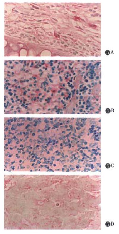
Figure 5 Immunohistochemical analysis of DTH reaction.
A.Ear: Most of the macrophages in subepidermal interstitial tissue and in the local capillary were obviously positive in their cytoplasm for the staining. S. Spleen: There were plenty of reticular-macrophage in red medulla. A few slightly positive cells, however, could be seen scattered there. C. The negative result was used by anti-GST F(ab’)2. D. Liver: The cytoplasm of Kupffer cells were slightly stained by the dyes.
- Citation: Guo BY, Zhang SY, Mukaida N, Harada A, Kuno K, Wang JB, Sun SH, Matshshima K. CCR5 gene expression in fulminant hepatitis and DTH in mice. World J Gastroenterol 1998; 4(1): 14-18
- URL: https://www.wjgnet.com/1007-9327/full/v4/i1/14.htm
- DOI: https://dx.doi.org/10.3748/wjg.v4.i1.14


