Copyright
©The Author(s) 2025.
World J Gastroenterol. Mar 7, 2025; 31(9): 98027
Published online Mar 7, 2025. doi: 10.3748/wjg.v31.i9.98027
Published online Mar 7, 2025. doi: 10.3748/wjg.v31.i9.98027
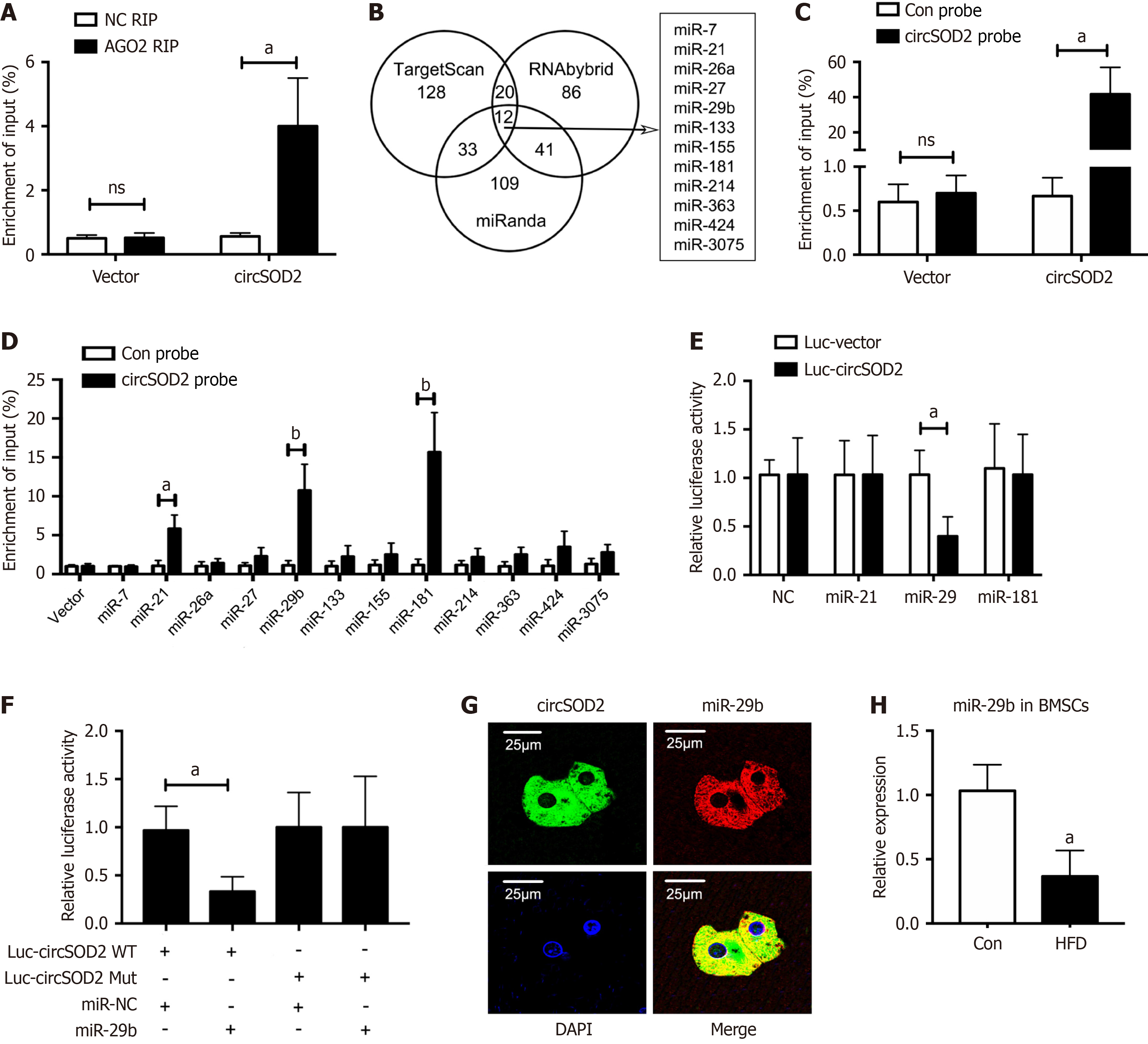
Figure 5 circSOD2 sponges miR-29b.
A: Reverse transcription-quantitative polymerase chain reaction (qRT-PCR) analysis of argonaute-2-bound circSOD2 using RNA immunoprecipitation; B: Venn diagram illustrating 12 potential target miRNAs of circSOD2 through cross-referencing TargetScan, RNAbybrid, and miRanda databases; C: Quantification of the efficiency of the circSOD2 probe for RNA antisense purification analysis using qRT-PCR; D: Quantification of the circSOD2-bound miRNAs using qRT-PCR; E: Relative luciferase activity of circSOD2 Luciferase reporter plasmids co-transfected with candidate miRNA mimics in HEK-293T cells; F: Quantification of relative luciferase activity using dual luciferase reporter assay after co-transfection of Luc-circSOD2 WT or Mut with miR-29b mimic or negative control in HEK-293T cells; G: Fluorescence in situ hybridization assay images demonstrating co-localization of circSOD2 and miR-29b in the cytoplasm; H: Decreased expression of miR-29b in bone marrow mesenchymal stem cells from high-fat diet mice (n = 5). aP < 0.05; bP < 0.01; NS: Not significant; NC: Negative control; AGO2: Analysis of argonaute-2; RIP: RNA immunoprecipitation; WT: Wildtype; Mut: Mutation; Con: Control.
- Citation: Li LP, Chen XY, Liu HB, Zhu Y, Xie MJ, Li YJ, Luo M, Albahde M, Zhang HY, Lou JY. Oxidative stress-induced circSOD2 inhibits osteogenesis through sponging miR-29b in metabolic-associated fatty liver disease. World J Gastroenterol 2025; 31(9): 98027
- URL: https://www.wjgnet.com/1007-9327/full/v31/i9/98027.htm
- DOI: https://dx.doi.org/10.3748/wjg.v31.i9.98027


