Copyright
©The Author(s) 2025.
World J Gastroenterol. Mar 7, 2025; 31(9): 102059
Published online Mar 7, 2025. doi: 10.3748/wjg.v31.i9.102059
Published online Mar 7, 2025. doi: 10.3748/wjg.v31.i9.102059
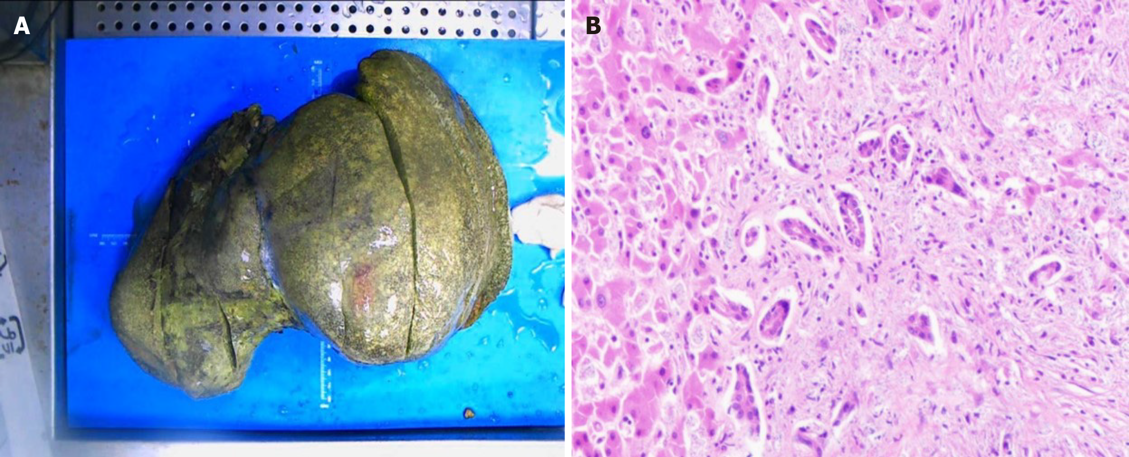
Figure 2 Pathological specimen.
A: Specimen of diseased liver (fixed in formalin), markedly enlarged in size (38 cm × 25 cm × 15 cm), dark in color, and hardened in texture, with a large number of yellow spots on the surface, and no obvious masses or nodules; B: Hematoxylin and eosin staining (100 ×). The normal hepatic lobular, structure is disrupted, with sub-massive necrosis of hepatic tissue, hyperplasia of small bile ducts, and a high level of lymphocytic infiltration.
- Citation: Li CX, Chen HD, Kashif S, Xie B, Luo J, Li T. Disseminated liver histoplasmosis in an immunocompetent individual cured by liver transplantation and voriconazole from China: A case report. World J Gastroenterol 2025; 31(9): 102059
- URL: https://www.wjgnet.com/1007-9327/full/v31/i9/102059.htm
- DOI: https://dx.doi.org/10.3748/wjg.v31.i9.102059


