Copyright
©The Author(s) 2025.
World J Gastroenterol. Feb 28, 2025; 31(8): 102071
Published online Feb 28, 2025. doi: 10.3748/wjg.v31.i8.102071
Published online Feb 28, 2025. doi: 10.3748/wjg.v31.i8.102071
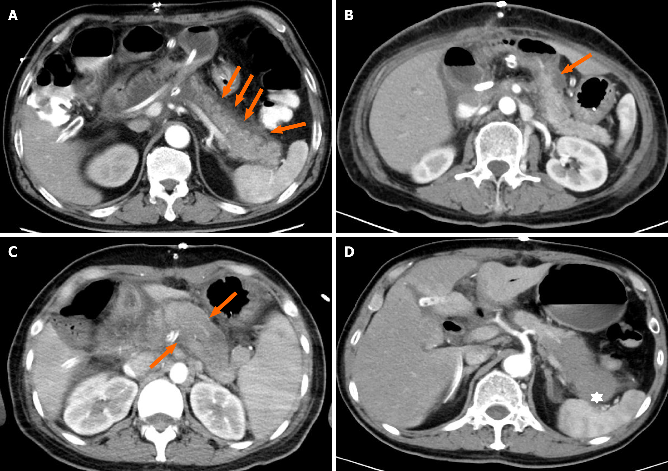
Figure 1 Several typical computed tomography changes of postpancreatectomy acute pancreatitis.
A: Peripancreatic fluid extravasation: Indicated by arrows as exudation belt; B: Residual peripancreatic fluid collection: Indicated by arrows as low-density fluid accumulation; C: Edema: Noticeable increase in the diameter of the pancreatic body and tail compared to preoperative dimensions; D: Necrotizing pancreatitis: Low-density shadow near the splenic hilum in the pancreatic tail.
- Citation: Ma JM, Wang PF, Yang LQ, Wang JK, Song JP, Li YM, Wen Y, Tang BJ, Wang XD. Machine learning model-based prediction of postpancreatectomy acute pancreatitis following pancreaticoduodenectomy: A retrospective cohort study. World J Gastroenterol 2025; 31(8): 102071
- URL: https://www.wjgnet.com/1007-9327/full/v31/i8/102071.htm
- DOI: https://dx.doi.org/10.3748/wjg.v31.i8.102071


