Copyright
©The Author(s) 2025.
World J Gastroenterol. Feb 28, 2025; 31(8): 100146
Published online Feb 28, 2025. doi: 10.3748/wjg.v31.i8.100146
Published online Feb 28, 2025. doi: 10.3748/wjg.v31.i8.100146
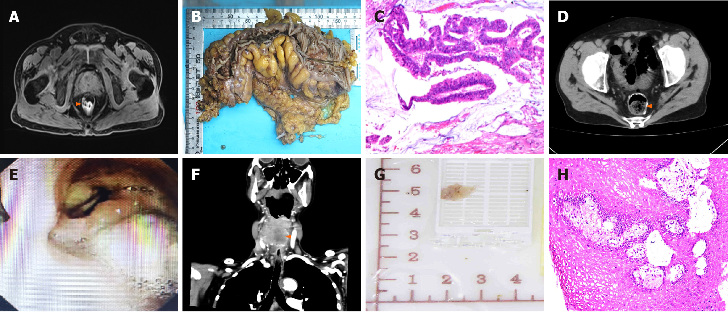
Figure 1 Main examination picture results of Case 1.
A: Abdominal magnetic resonance imaging revealed sigmoid colon wall thickening, highly suspicious for sigmoid colon carcinoma; B: Intraoperative gross specimen of colon carcinoma, mass size 15 cm × 1 cm × 0.5 cm; C: Colon carcinoma postoperative pathology (HE, × 100) results confirmed colon cancer, ulcerative type, presenting as a moderately differentiated tubular papillary adenocarcinoma. And was no evidence of peritoneal or lymph node involvement; D: Repeat computed tomography (CT) six months after surgery for colon carcinoma; E: Hypopharyngeal carcinoma preoperative laryngoscopy: A neoplasm with a rough surface was noted on the right lateral pharyngeal wall. This mass extended into the posterior pharyngeal wall and the right pyriform fossa, displacing the laryngopharynx to the left; F: Repeat CT six months after surgery for hypopharyngeal carcinoma; G: Intraoperative gross specimen of hypopharyngeal carcinoma, mass size 0.8 cm × 0.6 cm × 0.3 cm; H: Hypopharyngeal carcinoma postoperative pathology (HE, × 200).
- Citation: Bi XR, Zhao SY, Ma YQ, Duan XY, Hu TT, Bi LZ, Cai HY. Multiple primary cancers with gastrointestinal malignant tumors as the first manifestation: Three case reports and review of literature. World J Gastroenterol 2025; 31(8): 100146
- URL: https://www.wjgnet.com/1007-9327/full/v31/i8/100146.htm
- DOI: https://dx.doi.org/10.3748/wjg.v31.i8.100146


