Copyright
©The Author(s) 2025.
World J Gastroenterol. Feb 21, 2025; 31(7): 97599
Published online Feb 21, 2025. doi: 10.3748/wjg.v31.i7.97599
Published online Feb 21, 2025. doi: 10.3748/wjg.v31.i7.97599
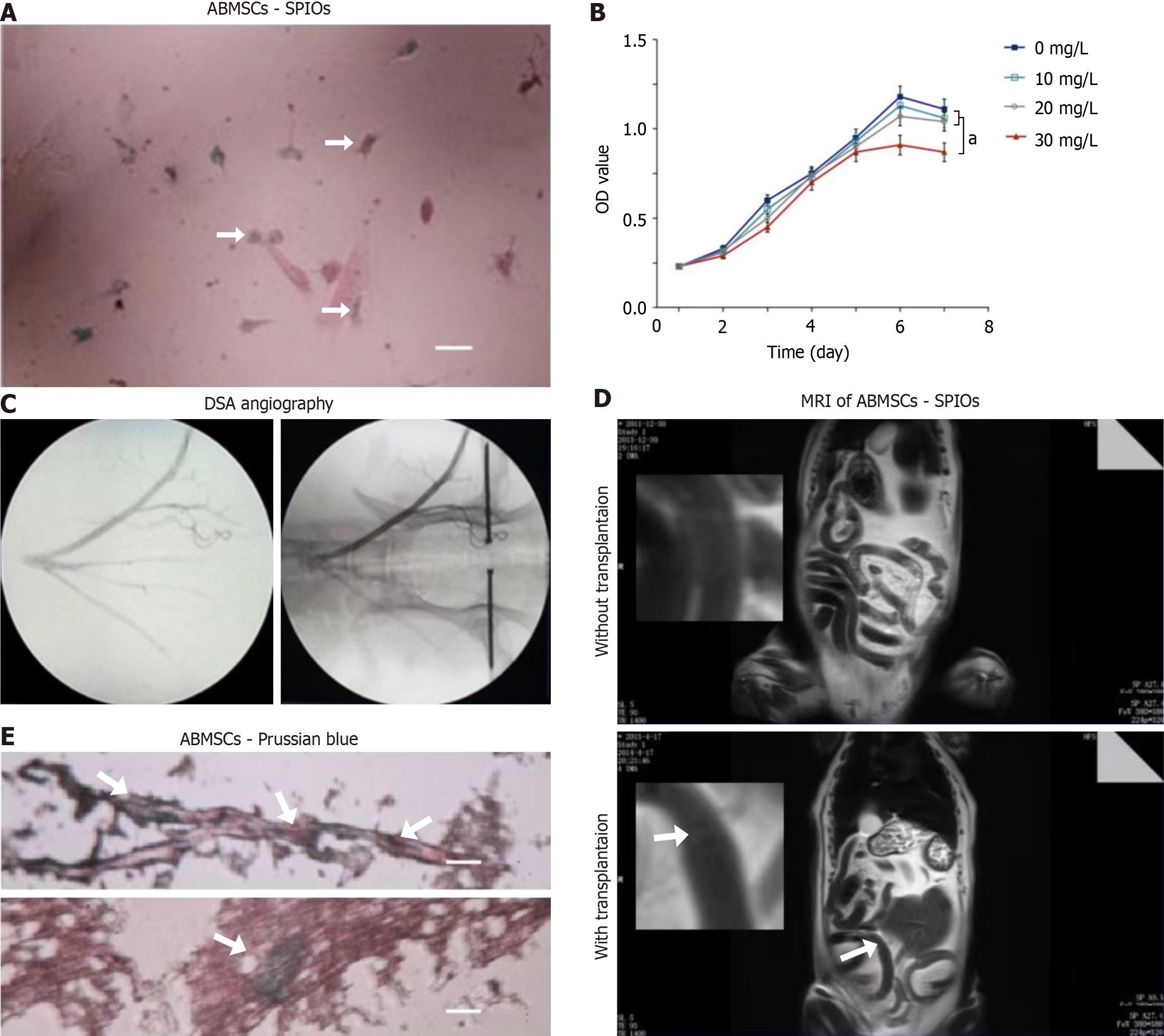
Figure 3 Transplantation and tracing for super paramagnetic iron oxide-labeled autologous bone marrow-derived mesenchymal stem cells in vivo.
A: Identification of cultured autologous bone marrow-derived mesenchymal stem cells (ABMSCs) labeled with super paramagnetic iron oxides (SPIOs) via Prussian blue staining and light microscopy (bars, 200 μm); B: The growth curve of ABMSCs labeled with different dose of SPIOs was determined by MTT assay. Each experiment was conducted in triplicate, and the values are reported as the mean ± SD; C: Abdominal aorta and mes-enteric artery branches were checked using femoral artery puncture and digital subtraction angiography imaging; D: Magnetic resonance imaging showed the results of T2 weighted image intensity comparison at the same layer; E: Observation of SPIO and Prussian blue–stained frozen sections via light microscopy. Bars, 100 μm. aP < 0.05 vs Blank group vs 30 mg/L group; SPIO: Super paramagnetic iron oxide; ABMSCs: Autologous bone marrow-derived mesenchymal stem cells; MRI: Magnetic resonance imaging.
- Citation: Sun GC, Xu WD, Yao H, Chen J, Chai RN. Protective effects of autologous bone marrow-derived mesenchymal stem cell transplantation on acute radioactive enteritis in Beagle dogs. World J Gastroenterol 2025; 31(7): 97599
- URL: https://www.wjgnet.com/1007-9327/full/v31/i7/97599.htm
- DOI: https://dx.doi.org/10.3748/wjg.v31.i7.97599


