Copyright
©The Author(s) 2025.
World J Gastroenterol. Feb 14, 2025; 31(6): 96782
Published online Feb 14, 2025. doi: 10.3748/wjg.v31.i6.96782
Published online Feb 14, 2025. doi: 10.3748/wjg.v31.i6.96782
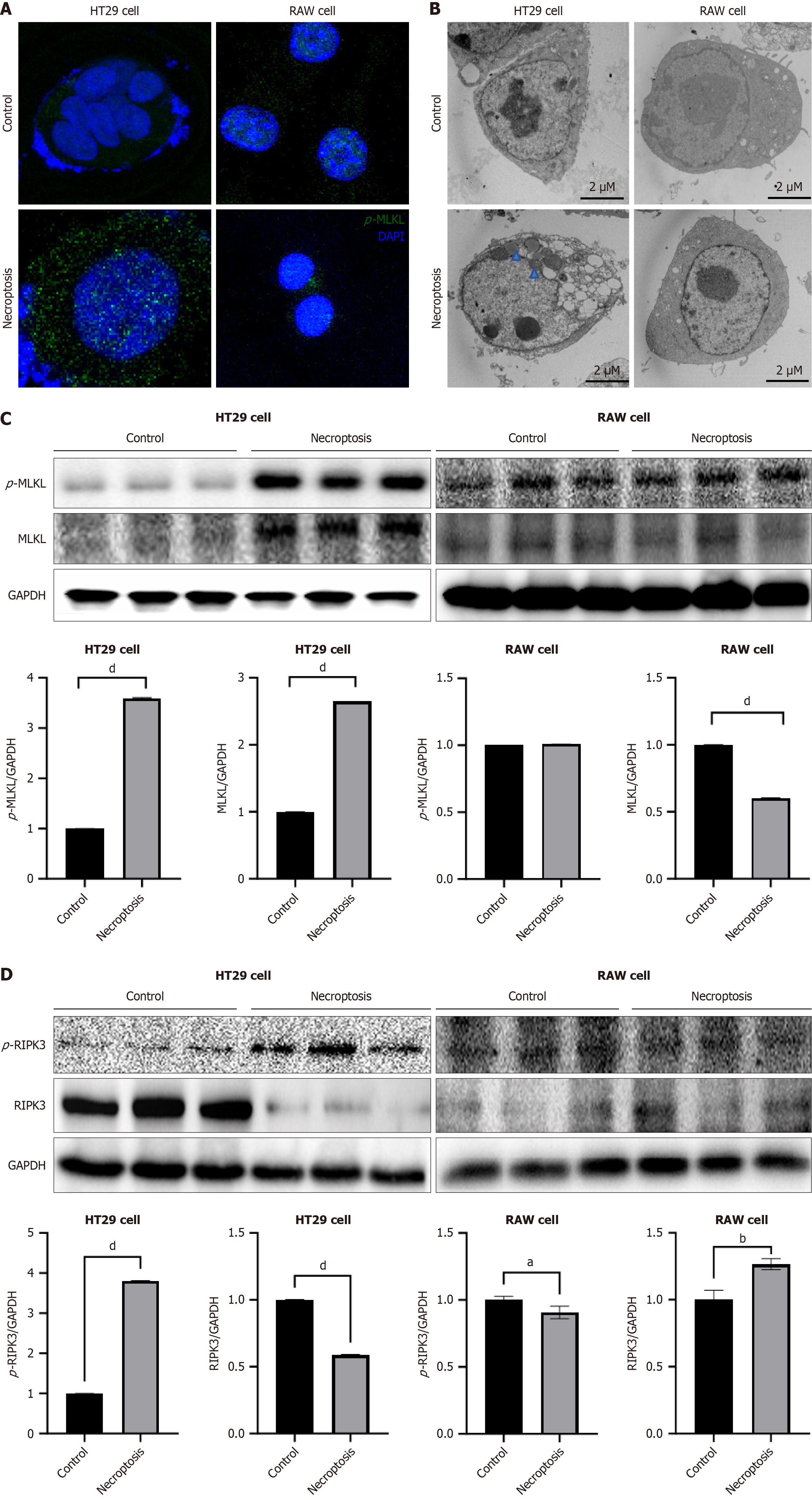
Figure 2 RAW cells did not phosphorylate mixed lineage kinase domain-like protein and avoided cell death after necroptosis stimuli.
A: Fixed and permeabilized HT29 and RAW 264.7 cells stained with 4’,6-diamidino-2-phenylindole were imaged by confocal microscopy before necroptosis induced [antibody: Phosphorylated-mixed lineage kinase domain-like protein (p-MLKL)]; B: Representative transmission electron microscopy images of HT29 and RAW 264.7 cells undergoing necroptosis for 24 hours. The blue arrow represents mitochondria, whereas the black arrow represents the plasma membrane (Scale bars: 2 μm); C: Western blot analysis of p-MLKL and MLKL protein expression in HT29 and RAW 264.7 cells before necroptosis; D: Western blot analysis of phosphorylated-receptor-interacting serine/threonine kinase 3 (RIPK3) and RIPK3 protein expression in HT29 and RAW 264.7 cells after necroptosis. aP < 0.05. bP < 0.01. dP < 0.0001. p-MLKL: Phosphorylated-mixed lineage kinase domain-like protein; DAPI: 4’,6-Diamidino-2-phenylindole dihydrochloride; MLKL: Mixed lineage kinase domain-like protein; RIPK3: Receptor-interacting serine/threonine kinase 3; p-RIPK3: Phosphorylated-receptor-interacting serine/threonine kinase 3; GAPDH: Glyceraldehyde 3-phosphate dehydrogenase.
- Citation: Xuan Yuan HN, Kim HS, Park GR, Ryu JE, Kim JE, Kang IY, Kim HY, Lee SM, Oh JH, Yoon EL, Jun DW. Adenosine triphosphate-binding pocket inhibitor for mixed lineage kinase domain-like protein attenuated alcoholic liver disease via necroptosis-independent pathway. World J Gastroenterol 2025; 31(6): 96782
- URL: https://www.wjgnet.com/1007-9327/full/v31/i6/96782.htm
- DOI: https://dx.doi.org/10.3748/wjg.v31.i6.96782


