Copyright
©The Author(s) 2025.
World J Gastroenterol. Feb 14, 2025; 31(6): 96782
Published online Feb 14, 2025. doi: 10.3748/wjg.v31.i6.96782
Published online Feb 14, 2025. doi: 10.3748/wjg.v31.i6.96782
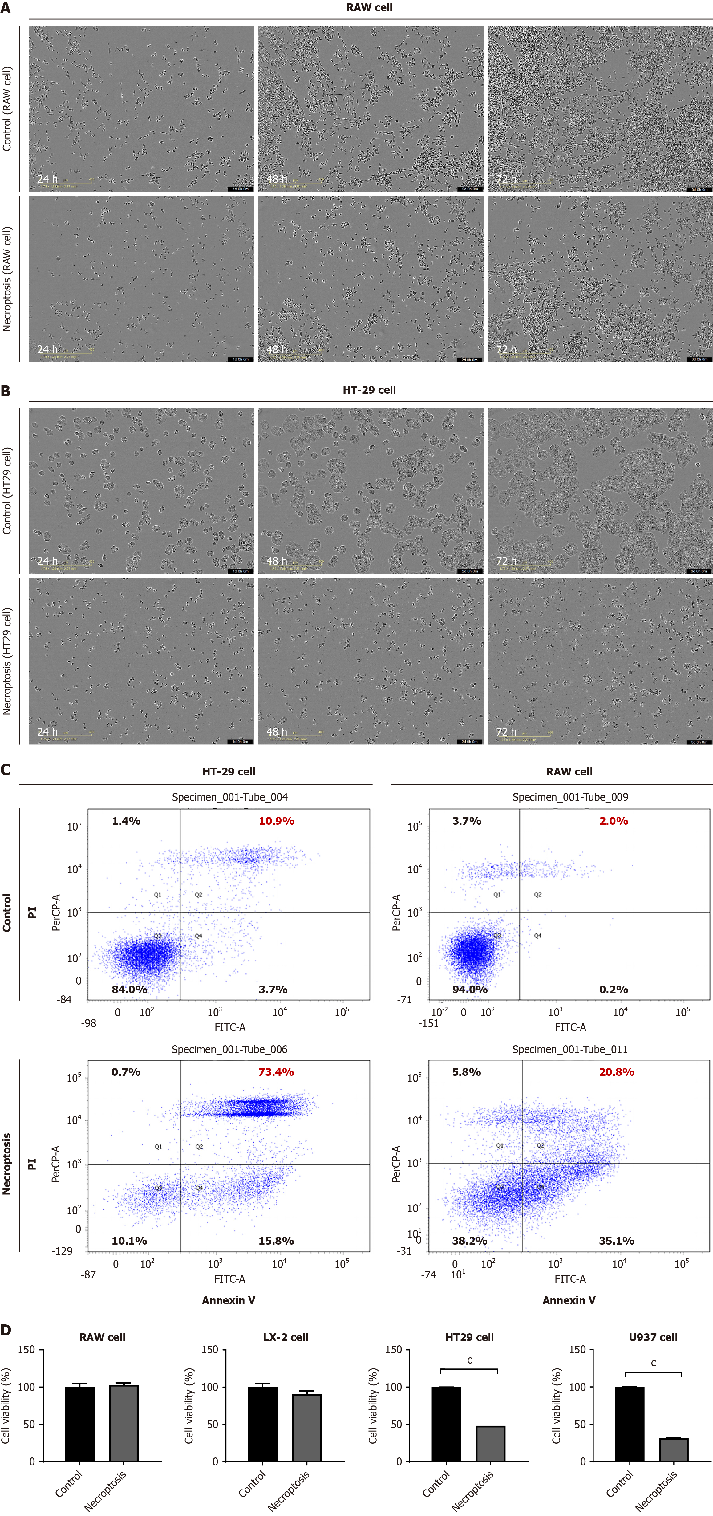
Figure 1 Cell death after necroptosis stimuli differed according to cell type.
A: After inducing necroptosis in RAW 264.7 cells, cell viability was determined using Incucyte assay; B: After inducing necroptosis in HT29 cells, cell viability was detected using Incucyte assay; C: Flow cytometry shows the ratio of necrosis induced in RAW 264.7 and HT9 cells after necroptosis induction; D: Cell viability was detected after necroptosis. Results are shown as mean. cP < 0.001. PI: Propidium iodide.
- Citation: Xuan Yuan HN, Kim HS, Park GR, Ryu JE, Kim JE, Kang IY, Kim HY, Lee SM, Oh JH, Yoon EL, Jun DW. Adenosine triphosphate-binding pocket inhibitor for mixed lineage kinase domain-like protein attenuated alcoholic liver disease via necroptosis-independent pathway. World J Gastroenterol 2025; 31(6): 96782
- URL: https://www.wjgnet.com/1007-9327/full/v31/i6/96782.htm
- DOI: https://dx.doi.org/10.3748/wjg.v31.i6.96782


