Copyright
©The Author(s) 2025.
World J Gastroenterol. Jan 28, 2025; 31(4): 101288
Published online Jan 28, 2025. doi: 10.3748/wjg.v31.i4.101288
Published online Jan 28, 2025. doi: 10.3748/wjg.v31.i4.101288
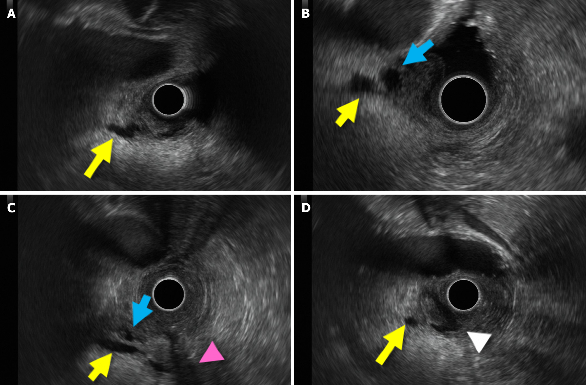
Figure 2 Utility of gel in endoscopic ultrasound for hepatobiliary and pancreatic visualization.
A: Clear depiction of the main pancreatic duct near the papilla (yellow arrow); B: The confluence of the main pancreatic duct and bile duct (blue arrow); C: Pancreatic and bile ducts relation to the duodenal muscle layer (magenta arrowhead); D: The pancreatic duct and the ampulla of Vater (white arrowhead) are depicted.
- Citation: Sato H, Kawabata H, Fujiya M. Gel immersion in endoscopy: Exploring potential applications. World J Gastroenterol 2025; 31(4): 101288
- URL: https://www.wjgnet.com/1007-9327/full/v31/i4/101288.htm
- DOI: https://dx.doi.org/10.3748/wjg.v31.i4.101288


