Copyright
©The Author(s) 2025.
World J Gastroenterol. Jan 14, 2025; 31(2): 98031
Published online Jan 14, 2025. doi: 10.3748/wjg.v31.i2.98031
Published online Jan 14, 2025. doi: 10.3748/wjg.v31.i2.98031
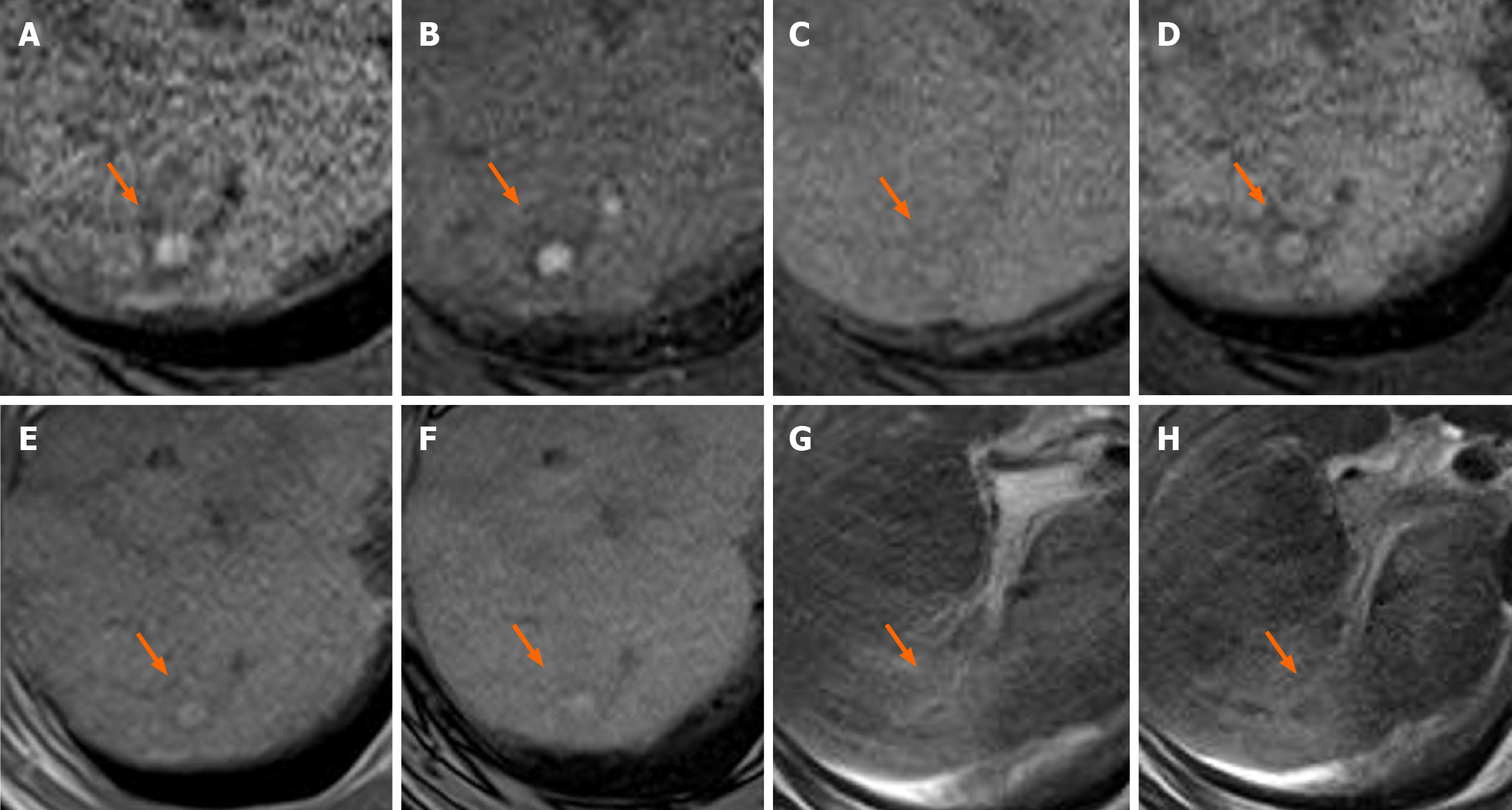
Figure 3 A 65-year-old man with alcoholic liver cirrhosis (patient No.
8). A and B: T1-weighted gradient-echo (GRE) magnetic resonance (MR) (A: In-phase; B: Opposed-phase) image shows a small and high-signal-intensity nodule (arrow) in segment 7 of the liver; C: Pre-contrast T1-weighted GRE MR imaging (MRI) shows iso-slightly high-intensity nodule (arrow); D and E: Gadoxetic acid-enhanced T1-weighted GRE MRI obtained during the arterial-and portal venous phase reveal a nodule with arterial phase hyperenhancement (arrow) and without washout (arrow); F: The hepatobiliary phase of the gadoxetic acid-enhanced T1-weighted GRE MRI shows homogeneous high-intense uptake (arrow); G: T2*-weighted GRE MRI shows iso-intensity nodule (arrow); H: The superparamagnetic iron oxide (SPIO)-enhanced T2*-weighted GRE MRI shows the lesion as a low-signal intensity nodule (arrow) with SPIO uptake compared with the surrounding liver parenchyma.
- Citation: Urase A, Tsurusaki M, Kozuki R, Kono A, Sofue K, Ishii K. Imaging characteristics of hypervascular focal nodular hyperplasia-like lesions in patients with chronic alcoholic liver disease. World J Gastroenterol 2025; 31(2): 98031
- URL: https://www.wjgnet.com/1007-9327/full/v31/i2/98031.htm
- DOI: https://dx.doi.org/10.3748/wjg.v31.i2.98031


