Copyright
©The Author(s) 2025.
World J Gastroenterol. May 7, 2025; 31(17): 105347
Published online May 7, 2025. doi: 10.3748/wjg.v31.i17.105347
Published online May 7, 2025. doi: 10.3748/wjg.v31.i17.105347
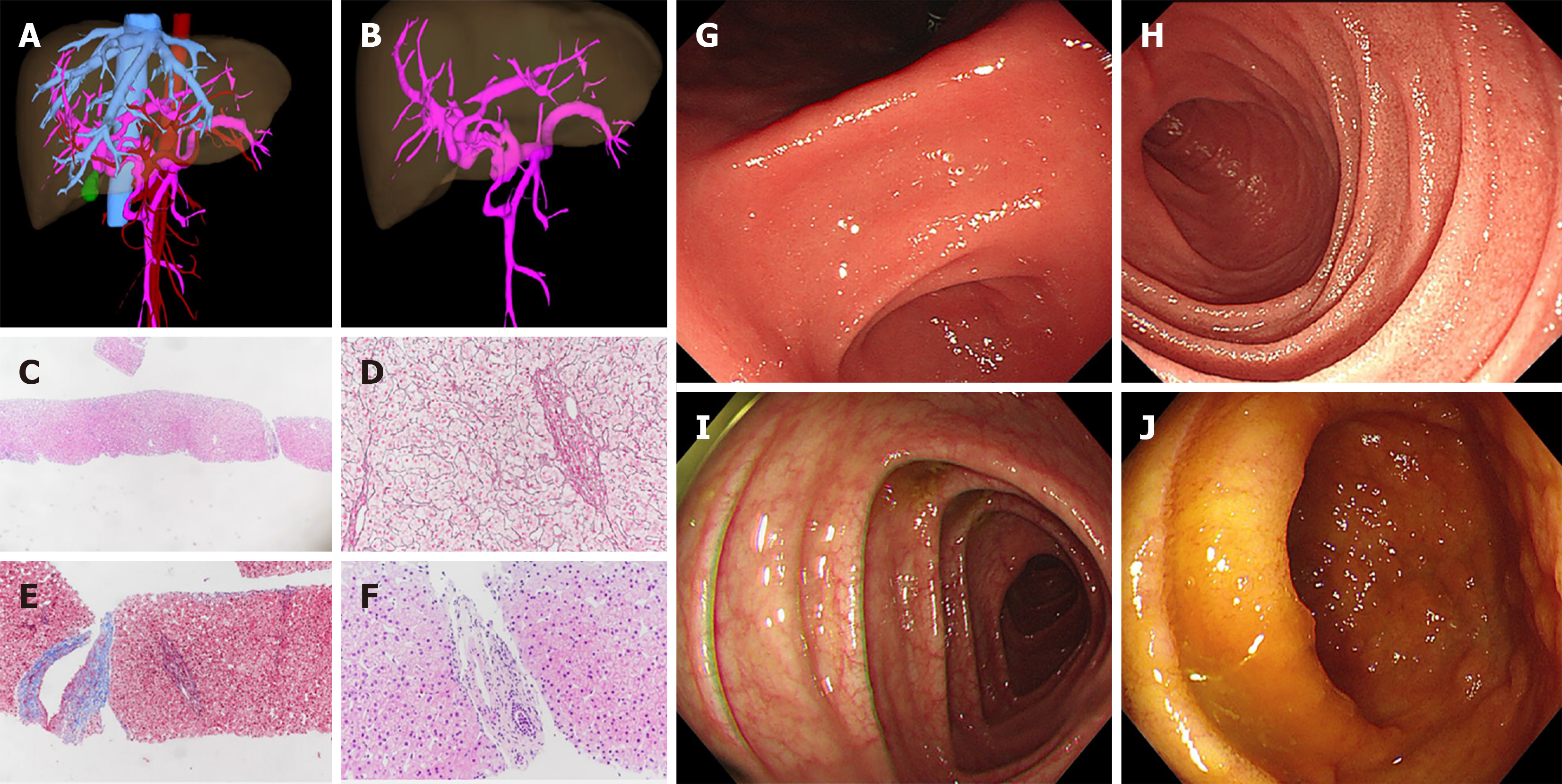
Figure 2 Imaging presentation about the liver and gastrointestinal tract.
A and B: Abdominal enhanced computed tomography by three-dimensional reconstruction indicating cavernous transformation of portal vein; C-F: Liver histology suggesting porto-sinusoidal vascular disease. The discernable lobular architecture (C: Hematoxylin and eosin staining, 200 ×), stenosis or disappearance of portal veins in portal areas (D: Reticular staining, 400 ×), herniated portal veins into the liver parenchyma, and smooth muscle proliferation in portal areas (E: Masson-trichrome staining, 200 ×), inflammatory cell infiltration not obvious in the portal areas (F: Hematoxylin and eosin staining, 400 ×); G and H: Upper and lower endoscopies showed no obvious abnormalities in the gastric and duodenal mucosa; I and J: No obvious abnormalities in the ileal and colonic mucosa except for the slight edema of the colon wall.
- Citation: Tian QJ, Zhang LJ, Zhang Q, Liu FC, Xie M, Cai JZ, Rao W. Protein-losing enteropathy and multiple vasculature dysplasia in LZTR1-related Noonan syndrome: A case report and review of literature. World J Gastroenterol 2025; 31(17): 105347
- URL: https://www.wjgnet.com/1007-9327/full/v31/i17/105347.htm
- DOI: https://dx.doi.org/10.3748/wjg.v31.i17.105347


