Copyright
©The Author(s) 2025.
World J Gastroenterol. Apr 14, 2025; 31(14): 104975
Published online Apr 14, 2025. doi: 10.3748/wjg.v31.i14.104975
Published online Apr 14, 2025. doi: 10.3748/wjg.v31.i14.104975
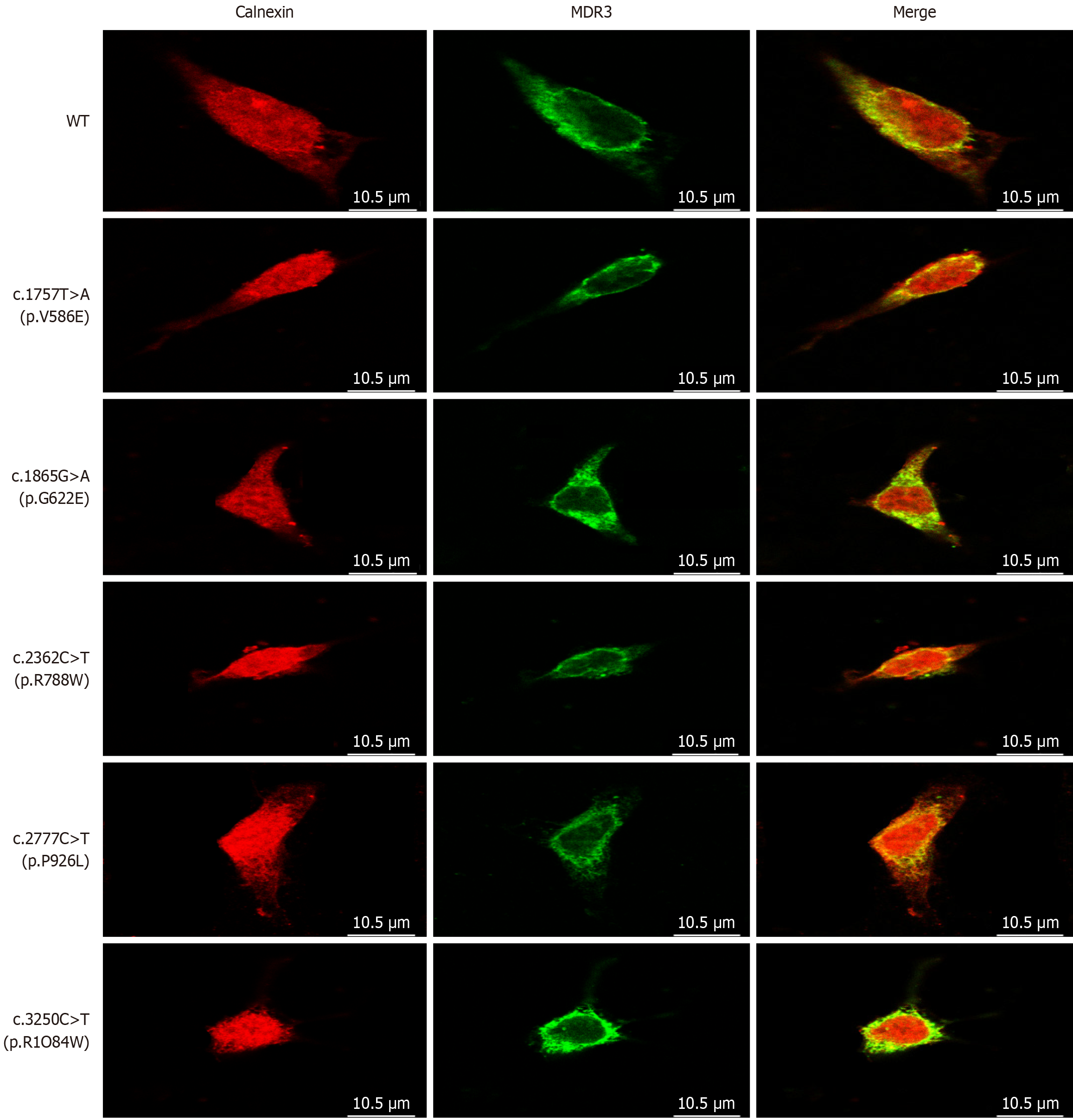
Figure 4 Confocal microscopy of cellular localization of multidrug resistance protein 3.
HEK293 cells expressing the wild-type and mutant ATP-binding cassette subfamily B member 4 alleles were labeled with anti- multidrug resistance protein 3 (red) and anti-calnexin (green) antibodies, the yellow color indicates the coexistence of the two proteins. Followed by fluorescence-conjugated secondary antibodies, and observed using laser scanning confocal microscopy. Bars = 10.5 μm. MDR3: Multidrug resistance protein 3; WT: Wild-type.
- Citation: Weng YH, Zheng YF, Yin DD, Xiong QF, Li JL, Li SX, Chen W, Yang YF. Clinical, genetic and functional perspectives on ATP-binding cassette subfamily B member 4 variants in five cholestasis adults. World J Gastroenterol 2025; 31(14): 104975
- URL: https://www.wjgnet.com/1007-9327/full/v31/i14/104975.htm
- DOI: https://dx.doi.org/10.3748/wjg.v31.i14.104975


