Copyright
©The Author(s) 2025.
World J Gastroenterol. Apr 7, 2025; 31(13): 102527
Published online Apr 7, 2025. doi: 10.3748/wjg.v31.i13.102527
Published online Apr 7, 2025. doi: 10.3748/wjg.v31.i13.102527
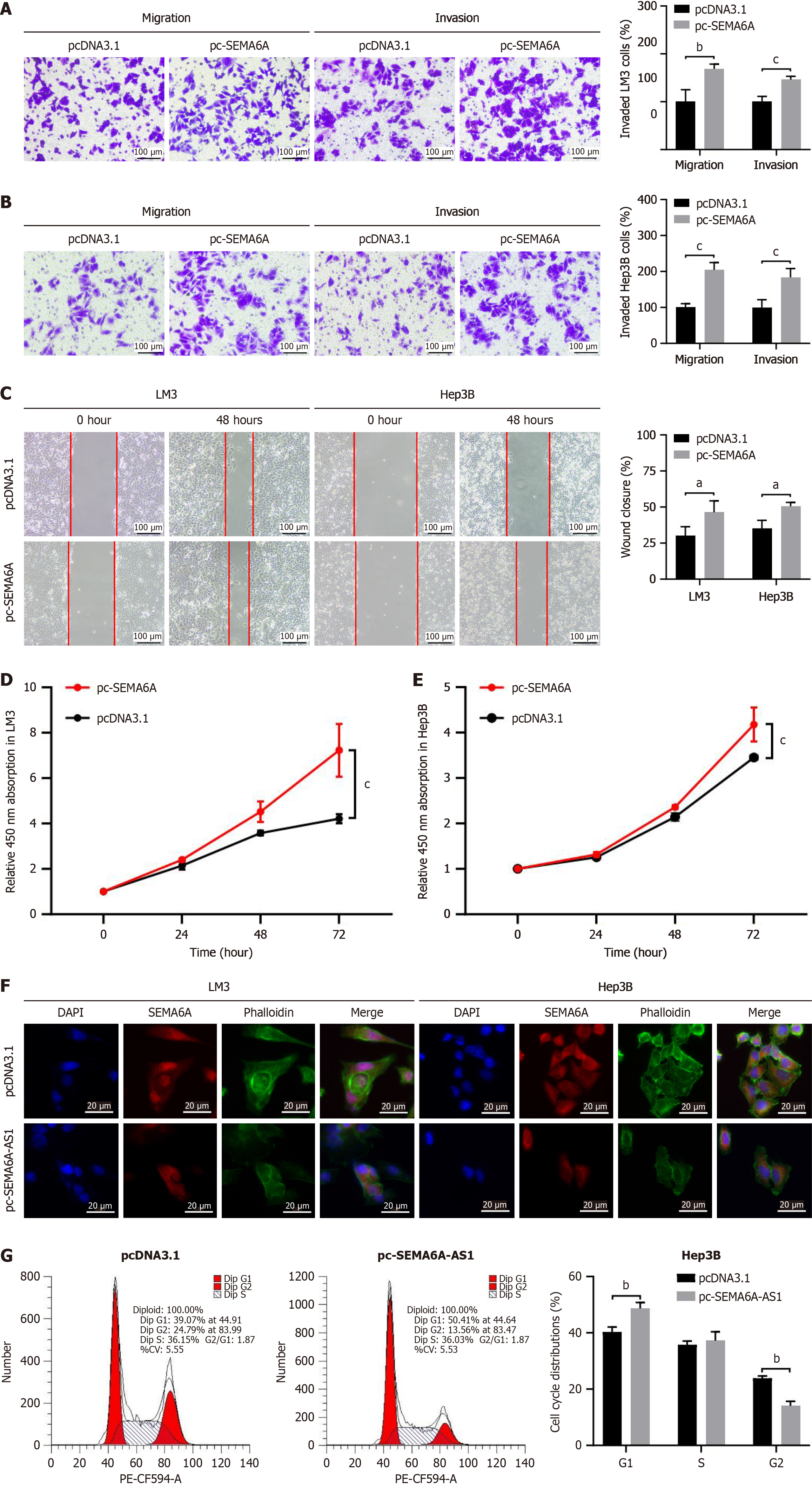
Figure 5 Semaphorin 6A promotes cell proliferation, migration and invasion via regulation of actin cytoskeleton.
A and B: Transwell assay with or without Matrigel were visualized (left panel) and quantified (right panel) in pc-semaphorin 6A (pc-SEMA6A) or pcDNA3.1 plasmid stably transfected LM3 (A) and Hep3B (B) cell lines to analyze cell invasion or migration capability; C: Wound healing assays in pc-SEMA6A or pcDNA3.1 plasmid stably transfected LM3 and Hep3B cell lines were visualized (left panel) and quantified (right panel) to assess cell migration ability; D: Cell proliferation in Hep3B cells stably transfected with pc-SEMA6A or pcDNA3.1 plasmid were analyzed by Cell Counting Kit-8 assay; E: Cell proliferation in LM3 cells stably transfected with pc-SEMA6A or pcDNA3.1 plasmid were analyzed by Cell Counting Kit-8 assay; F: Fluorescence microscopy imaging of SEMA6A (red), actin filaments stained with phalloidin (green) and nuclei stained with DAPI (blue) in LM3 and Hep3B cell lines (Scale bars: 20 μm); G: Flow cytometry assay was performed to analyze the cell cycle progression of pc-SEMA6A-AS1 or pcDNA3.1 plasmid stably transfected Hep3B cells. aP < 0.05, bP < 0.01, cP < 0.001. SEMA6A: Semaphorin 6A.
- Citation: Yu SM, Zhang M, Li SL, Pei SY, Wu L, Hu XW, Duan YK. Long noncoding RNA semaphorin 6A-antisense RNA 1 reduces hepatocellular carcinoma by promoting semaphorin 6A mRNA degradation. World J Gastroenterol 2025; 31(13): 102527
- URL: https://www.wjgnet.com/1007-9327/full/v31/i13/102527.htm
- DOI: https://dx.doi.org/10.3748/wjg.v31.i13.102527


