Copyright
©The Author(s) 2025.
World J Gastroenterol. Mar 28, 2025; 31(12): 103952
Published online Mar 28, 2025. doi: 10.3748/wjg.v31.i12.103952
Published online Mar 28, 2025. doi: 10.3748/wjg.v31.i12.103952
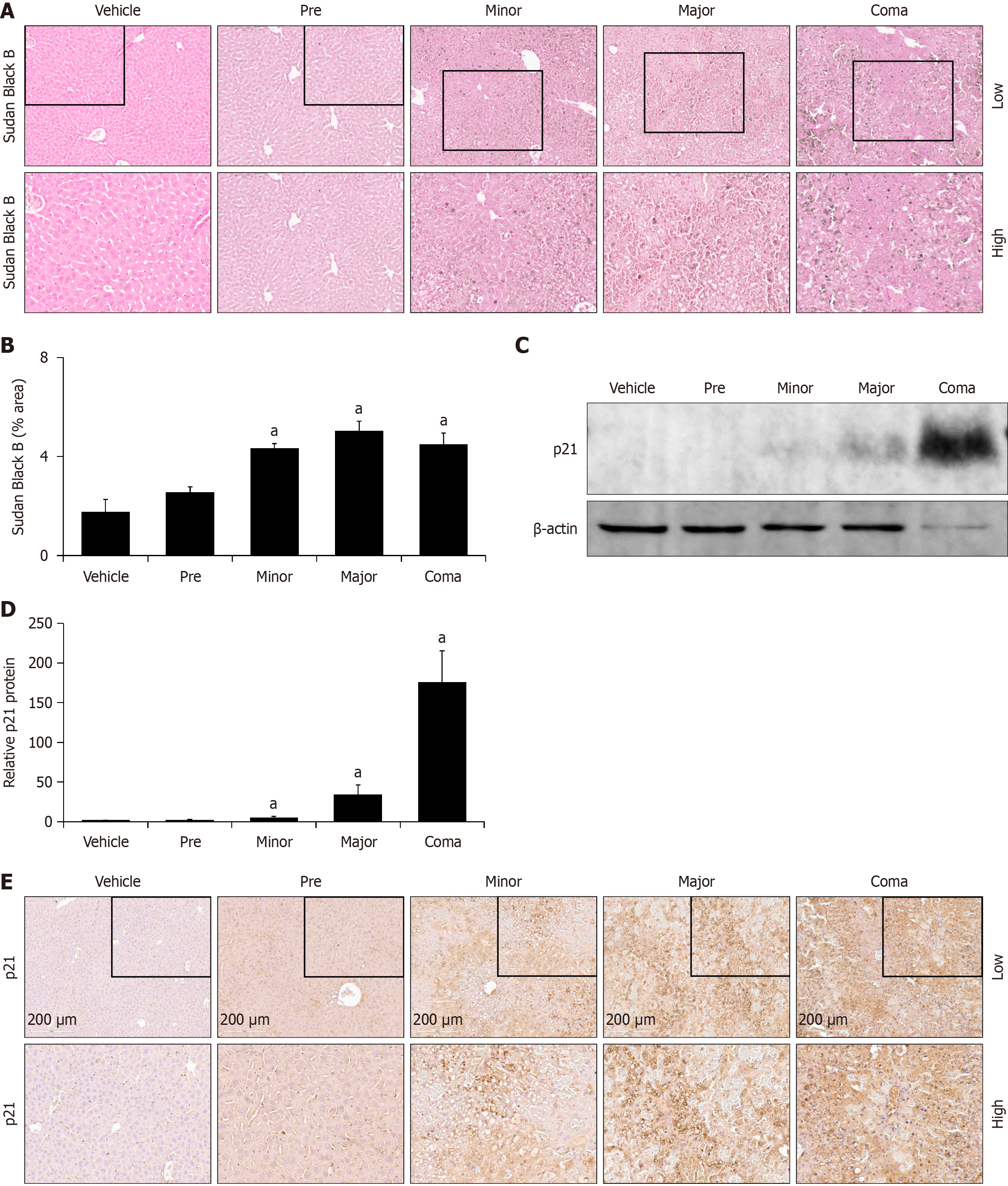
Figure 7 Hepatocellular senescence is increased in response to azoxymethane toxicity.
A: Representative Sudan Black B histochemistry images in liver from vehicle and azoxymethane (AOM)- treated mice at the pre, minor, major and coma time points; B: Liver Sudan Black B staining in mice treated with vehicle or AOM at pre, minor, major and coma time points reported in percent area; C and D: Representative immunoblot and quantification of relative p21 protein from liver homogenates of vehicle- and AOM-treated mice at the pre, minor, major and coma time points. β-actin was used as a protein loading control; E: Representative immunohistochemistry images stained for p21 in livers from vehicle- and AOM-treated mice at the pre, minor, major and coma time points. n ≥ 3 for each group for all analyses. aP < 0.05 compared to vehicle-treated mice.
- Citation: Bhattarai SM, Jhawer A, Frampton G, Troyanovskaya E, DeMorrow S, McMillin M. Characterization of hepatic pathology during azoxymethane-induced acute liver failure. World J Gastroenterol 2025; 31(12): 103952
- URL: https://www.wjgnet.com/1007-9327/full/v31/i12/103952.htm
- DOI: https://dx.doi.org/10.3748/wjg.v31.i12.103952


