Copyright
©The Author(s) 2025.
World J Gastroenterol. Mar 28, 2025; 31(12): 100855
Published online Mar 28, 2025. doi: 10.3748/wjg.v31.i12.100855
Published online Mar 28, 2025. doi: 10.3748/wjg.v31.i12.100855
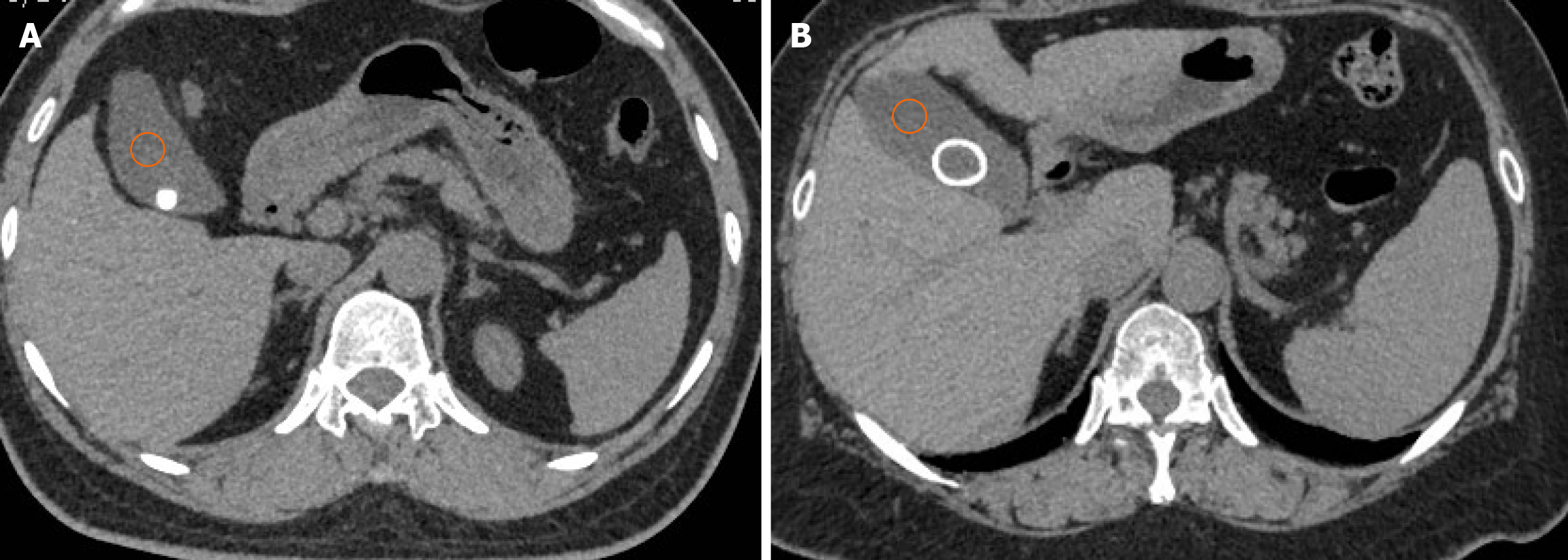
Figure 3 Non-enhanced CT scans.
A: The HU value measurement of homogeneous gallbladder stones; B: The HU value measurement of heterogeneous gallbladder stones. Three 2 cm2 circular regions of interest were manually drawn at three different parts of segments VII and VIII of the right lobe of the liver and the middle third of the spleen. The orange circle is the measured bile gray value.
- Citation: Qiu C, Xiang YK, Hu H, Da XB, Li G, Zhang YY, Zhang HL, Zhang C, Yang YL. Characterization of gallbladder stones associated with occult pancreaticobiliary reflux using computed tomography. World J Gastroenterol 2025; 31(12): 100855
- URL: https://www.wjgnet.com/1007-9327/full/v31/i12/100855.htm
- DOI: https://dx.doi.org/10.3748/wjg.v31.i12.100855


