Copyright
©The Author(s) 2025.
World J Gastroenterol. Mar 21, 2025; 31(11): 102848
Published online Mar 21, 2025. doi: 10.3748/wjg.v31.i11.102848
Published online Mar 21, 2025. doi: 10.3748/wjg.v31.i11.102848
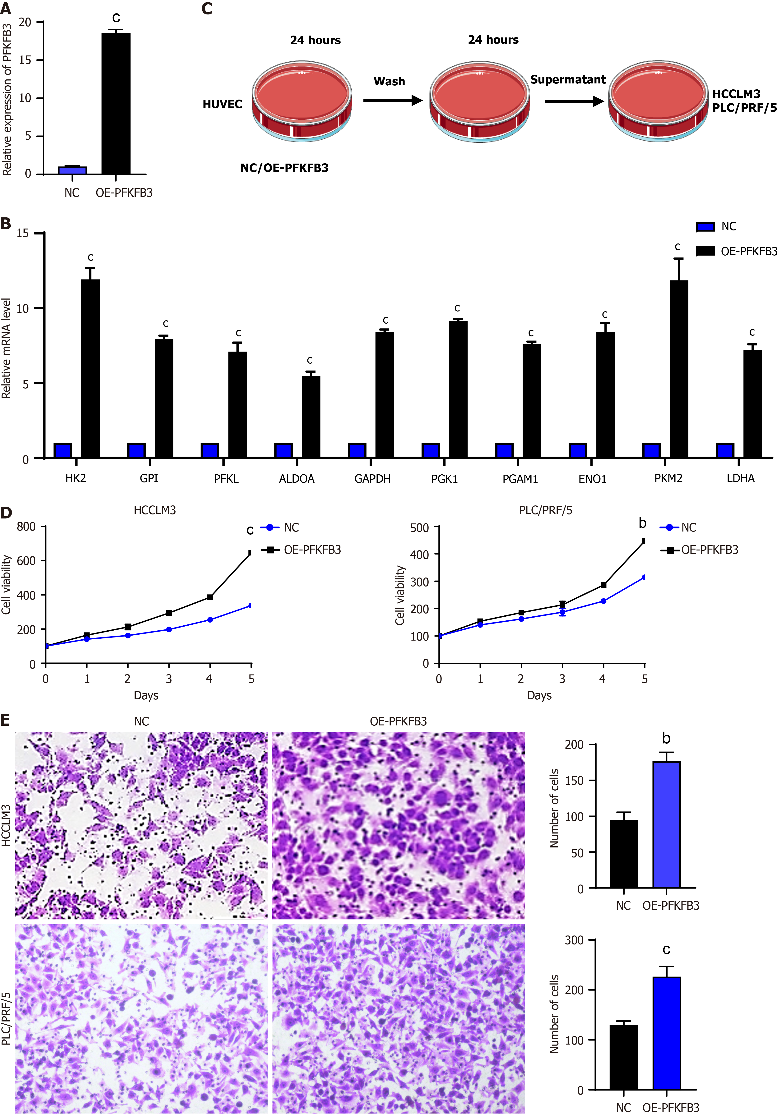
Figure 2 Increased proliferation and migration of liver cancer cells due to high glycolysis vascular endothelial cells.
A: Expression levels of 6-phosphofructo-2-kinase/fructose-2,6-biphosphatase 3 in human umbilical vein endothelial cells; B: Expression levels of key glycolytic enzymes in vascular endothelial cells (VECs) detected by quantitative real-time polymerase chain reaction; C: Schematic diagram of co-culture of VECs with liver cancer cells; D: Cell Counting Kit-8 proliferation assay of HCCLM3 and PLC/PRF/5 liver cancer cells; E: Transwell experiment of HCCLM3 and PLC/PRF/5. bP < 0.01, cP < 0.001. PFKFB3: 6-phosphofructo-2-kinase/fructose-2,6-biphosphatase 3; OE: Overexpression; HK2: Hexokinase 2; CPI: Cyclopropyl isomerase; PFKL: Phosphofructokinase liver type; ALDOA: Aldolase A; GAPDH: Glyceraldehyde-3-phosphate dehydrogenase; PGK1: Phosphoglycerate kinase 1; PGAM: Phosphoglycerate mutase; ENO: Enolase; PKM2: Pyruvate kinase M2; LDHA: Lactate dehydrogenase A.
- Citation: Wu Y, Xie BB, Zhang BL, Zhuang QX, Liu SW, Pan HM. Apatinib regulates the glycolysis of vascular endothelial cells through PI3K/AKT/PFKFB3 pathway in hepatocellular carcinoma. World J Gastroenterol 2025; 31(11): 102848
- URL: https://www.wjgnet.com/1007-9327/full/v31/i11/102848.htm
- DOI: https://dx.doi.org/10.3748/wjg.v31.i11.102848


