Copyright
©The Author(s) 2025.
World J Gastroenterol. Mar 21, 2025; 31(11): 102848
Published online Mar 21, 2025. doi: 10.3748/wjg.v31.i11.102848
Published online Mar 21, 2025. doi: 10.3748/wjg.v31.i11.102848
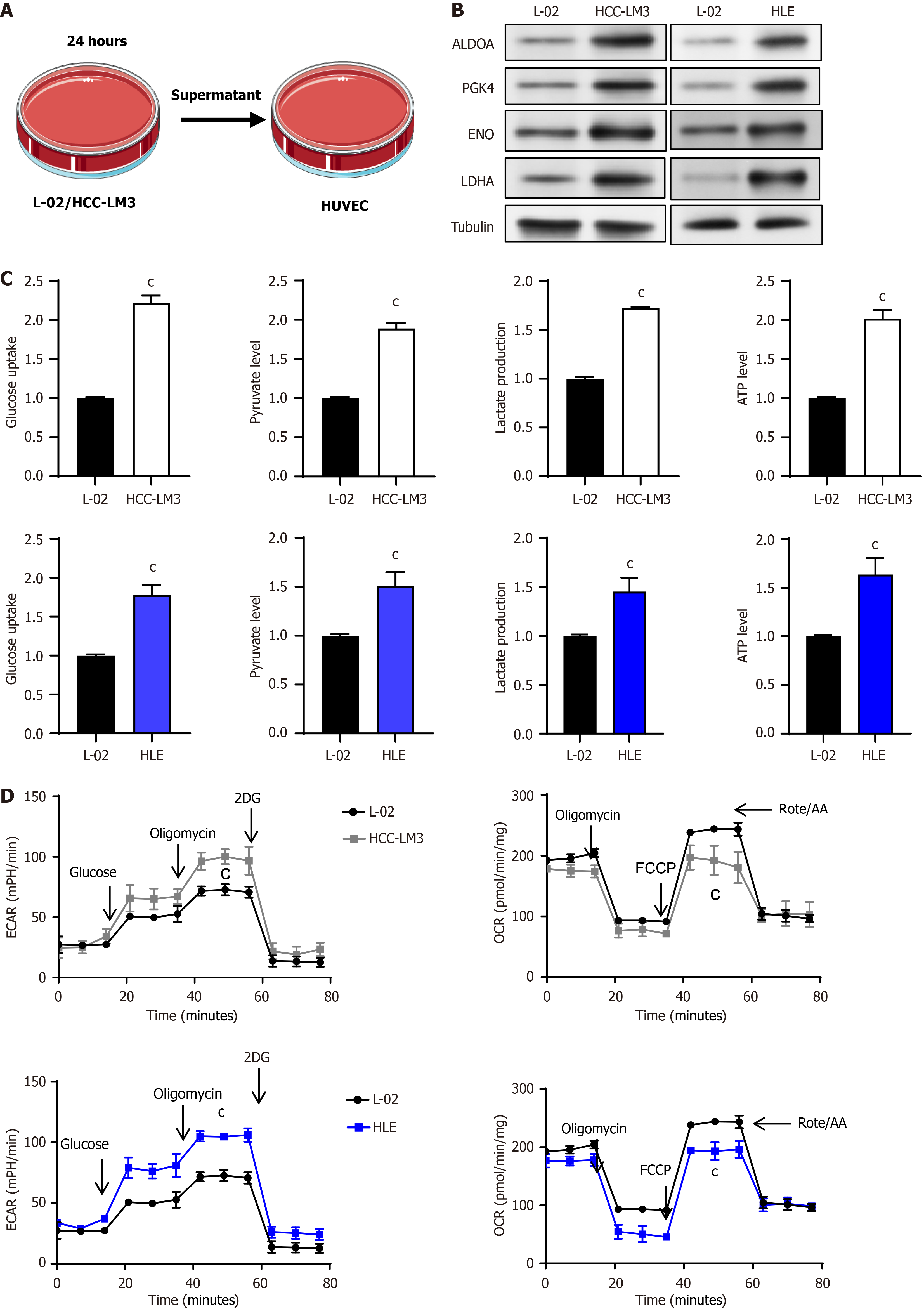
Figure 1 Enhanced glycolysis activity of vascular endothelial cells after co-culture with liver cancer cells.
A: Schematic diagram of co-culture of liver cells or liver cancer cells with vascular endothelial cells (VECs); B: Expression analysis of key glycolytic enzymes in VECs co-cultured with liver cells or liver cancer cells using Western blotting; C: Quantification of glucose consumption, pyruvate synthesis, lactate release, and adenosine triphosphate production during glycolysis in VECs; D: Evaluation of the glycolytic potential and oxidative phosphorylation efficiency of VECs when co-cultured with either liver cells or liver cancer cells, utilizing extracellular acidification rate and oxygen consumption rate. cP < 0.001. HUVEC: Human umbilical vein endothelial cells; ALDOA: Aldolase A, PGK: Phosphoglycerate kinase, ENO: Enolase, LDHA: Lactate dehydrogenase A; HCC: Hepatocellular carcinoma; ATP: Adenosine triphosphate; ECAR: Extracellular acidification rate; 2DG: 2-deoxy-D-glucose; OCR: Oxygen consumption rate; FCCP: Carbonylcyanide-p-trifluoromethoxyphenylhydrazone.
- Citation: Wu Y, Xie BB, Zhang BL, Zhuang QX, Liu SW, Pan HM. Apatinib regulates the glycolysis of vascular endothelial cells through PI3K/AKT/PFKFB3 pathway in hepatocellular carcinoma. World J Gastroenterol 2025; 31(11): 102848
- URL: https://www.wjgnet.com/1007-9327/full/v31/i11/102848.htm
- DOI: https://dx.doi.org/10.3748/wjg.v31.i11.102848


