Copyright
©The Author(s) 2024.
World J Gastroenterol. Mar 7, 2024; 30(9): 1189-1212
Published online Mar 7, 2024. doi: 10.3748/wjg.v30.i9.1189
Published online Mar 7, 2024. doi: 10.3748/wjg.v30.i9.1189
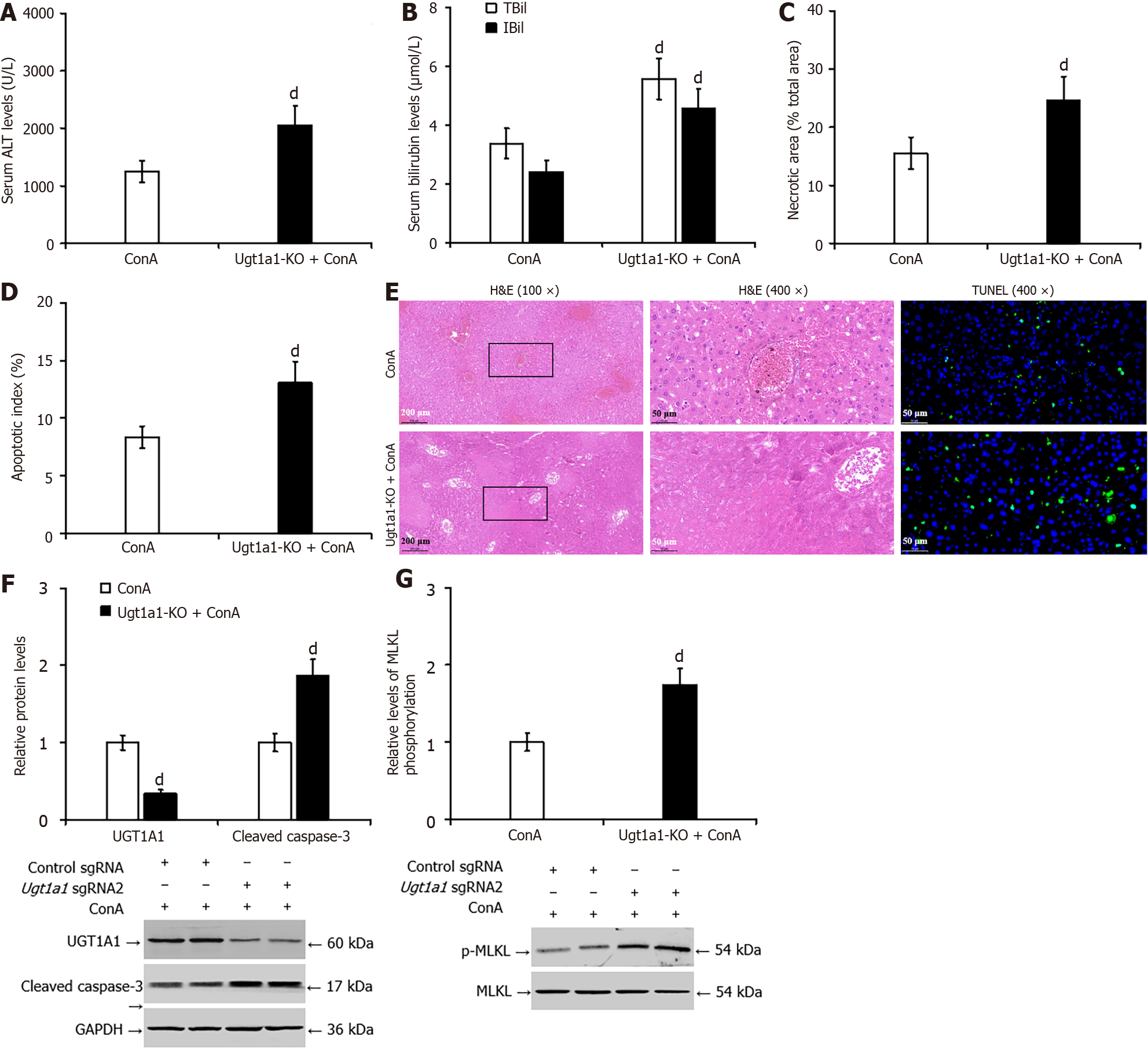
Figure 7 Knocking out uridine diphosphate glucuronosyltransferase 1A1 worsened concanavalin A-induced liver injury in mice.
A: The enzyme rate method was used to detect the serum level of alanine transaminase in mice; B: The diazo method was used to detect the serum level of total bilirubin and indirect bilirubin; C and E: Pathological analysis of liver tissue by hematoxylin and eosin staining; D and E: The terminal deoxynucleotidyl transferase-mediated deoxyuridine triphosphate-nick end labelling assay was used to measure hepatocyte apoptosis; F: Western blotting was used to detect the protein levels of uridine diphosphate glucuronosyltransferase 1A1 and cleaved caspase-3; G: Western blotting was used to detect the protein levels of phosphorylated mixed lineage kinase domain-like pseudokinase. dP < 0.01 vs the concanavalin A group. UGT1A1: Uridine diphosphate glucuronosyltransferase 1A1; ALT: Alanine transaminase; TBil: Total bilirubin; IBil: Indirect bilirubin; H&E: Hematoxylin and eosin; TUNEL: Transferase-mediated deoxyuridine triphosphate-nick end labelling; p-MLKL: Phosphorylated mixed lineage kinase domain-like pseudokinase; CCl4: Carbon tetrachloride; KO: Knockout; GAPDH: Glyceraldehyde 3 phosphate dehydrogenase.
- Citation: Jiang JL, Zhou YY, Zhong WW, Luo LY, Liu SY, Xie XY, Mu MY, Jiang ZG, Xue Y, Zhang J, He YH. Uridine diphosphate glucuronosyltransferase 1A1 prevents the progression of liver injury. World J Gastroenterol 2024; 30(9): 1189-1212
- URL: https://www.wjgnet.com/1007-9327/full/v30/i9/1189.htm
- DOI: https://dx.doi.org/10.3748/wjg.v30.i9.1189


