Copyright
©The Author(s) 2024.
World J Gastroenterol. Mar 7, 2024; 30(9): 1164-1176
Published online Mar 7, 2024. doi: 10.3748/wjg.v30.i9.1164
Published online Mar 7, 2024. doi: 10.3748/wjg.v30.i9.1164
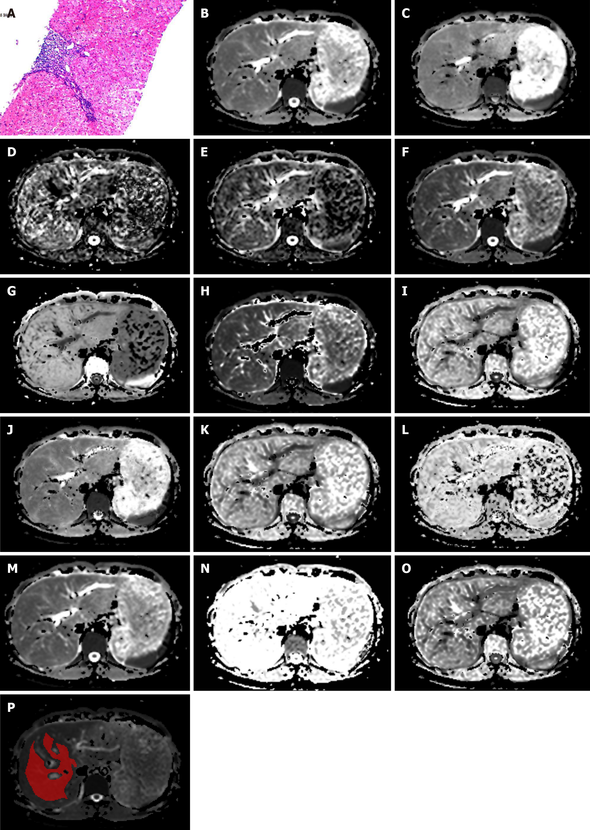
Figure 2 A 27-year-old female patient with hepatitis B virus for nine years.
The liver fibrosis stage was diagnosed as F1. A: Pathology image, H&E-stained samples (original magnification × 100) of right lobe of liver, shows portal fibrosis; B: Mono-apparent diffusion coefficient map; C-E: Intravoxel incoherent motion (IVIM) model-derived true diffusion coefficient, IVIM model-derived pseudo-diffusion coefficient, and IVIM model-derived perfusion fraction maps; F and G: Diffusion kurtosis imaging (DKI)-derived apparent diffusivity and DKI-derived excess kurtosis maps; H and I: Stretched exponential model (SEM)-derived distributed diffusion coefficient and SEM-derived intravoxel heterogeneity index maps; J-L: Fractional order calculus model-derived diffusion coefficient, fractional order calculus (FROC)-derived fractional order parameter, and FROC model-derived microstructural quantity maps; M-O: Continuous-time random-walk (CTRW) model-derived anomalous diffusion coefficient, CTRW model-derived temporal diffusion heterogeneity index, and CTRW model-derived spatial diffusion heterogeneity index maps; P: The region of interest placement in the liver parenchyma.
- Citation: Jiang YL, Li J, Zhang PF, Fan FX, Zou J, Yang P, Wang PF, Wang SY, Zhang J. Staging liver fibrosis with various diffusion-weighted magnetic resonance imaging models. World J Gastroenterol 2024; 30(9): 1164-1176
- URL: https://www.wjgnet.com/1007-9327/full/v30/i9/1164.htm
- DOI: https://dx.doi.org/10.3748/wjg.v30.i9.1164


