Copyright
©The Author(s) 2024.
World J Gastroenterol. Mar 7, 2024; 30(9): 1096-1107
Published online Mar 7, 2024. doi: 10.3748/wjg.v30.i9.1096
Published online Mar 7, 2024. doi: 10.3748/wjg.v30.i9.1096
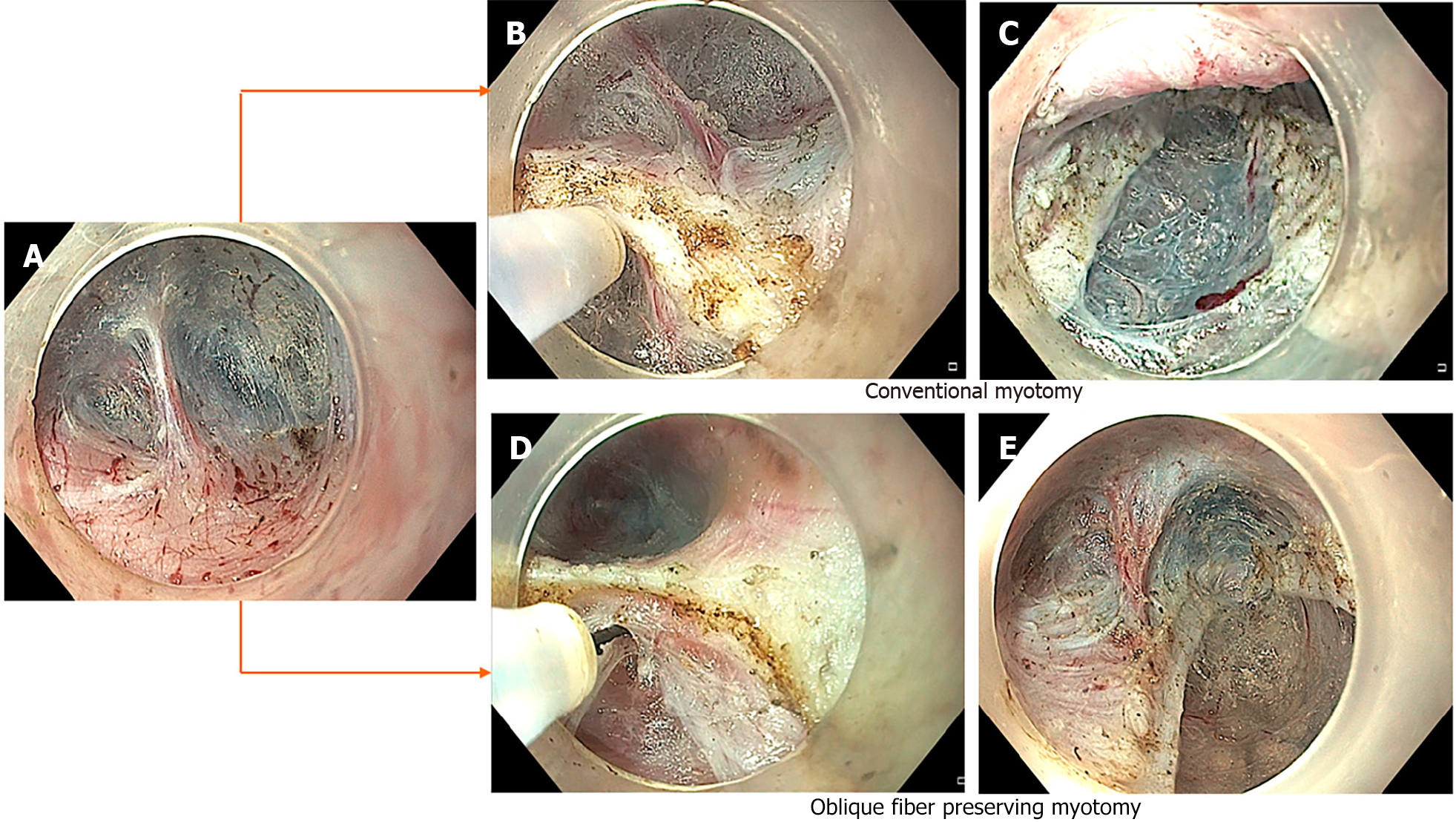
Figure 2 Conventional and sling fiber preservation technique of per-oral endoscopic myotomy.
A: Endoscopic image revealing second penetrating vessel along gastric side. Note that the sling fibers are located towards the left of penetrating vessel; B: Conventional myotomy performed along left side of the penetrating vessel to include sling fibers; C: Completion of conventional myotomy; D: Myotomy along the right side of the penetrating vessel to preserve sling fibers; E: Completion of myotomy (Note the preservation of sling fibers towards the left of second penetrating vessel).
- Citation: Nabi Z, Inavolu P, Duvvuru NR. Prediction, prevention and management of gastroesophageal reflux after per-oral endoscopic myotomy: An update. World J Gastroenterol 2024; 30(9): 1096-1107
- URL: https://www.wjgnet.com/1007-9327/full/v30/i9/1096.htm
- DOI: https://dx.doi.org/10.3748/wjg.v30.i9.1096


