Copyright
©The Author(s) 2024.
World J Gastroenterol. Mar 7, 2024; 30(9): 1043-1072
Published online Mar 7, 2024. doi: 10.3748/wjg.v30.i9.1043
Published online Mar 7, 2024. doi: 10.3748/wjg.v30.i9.1043
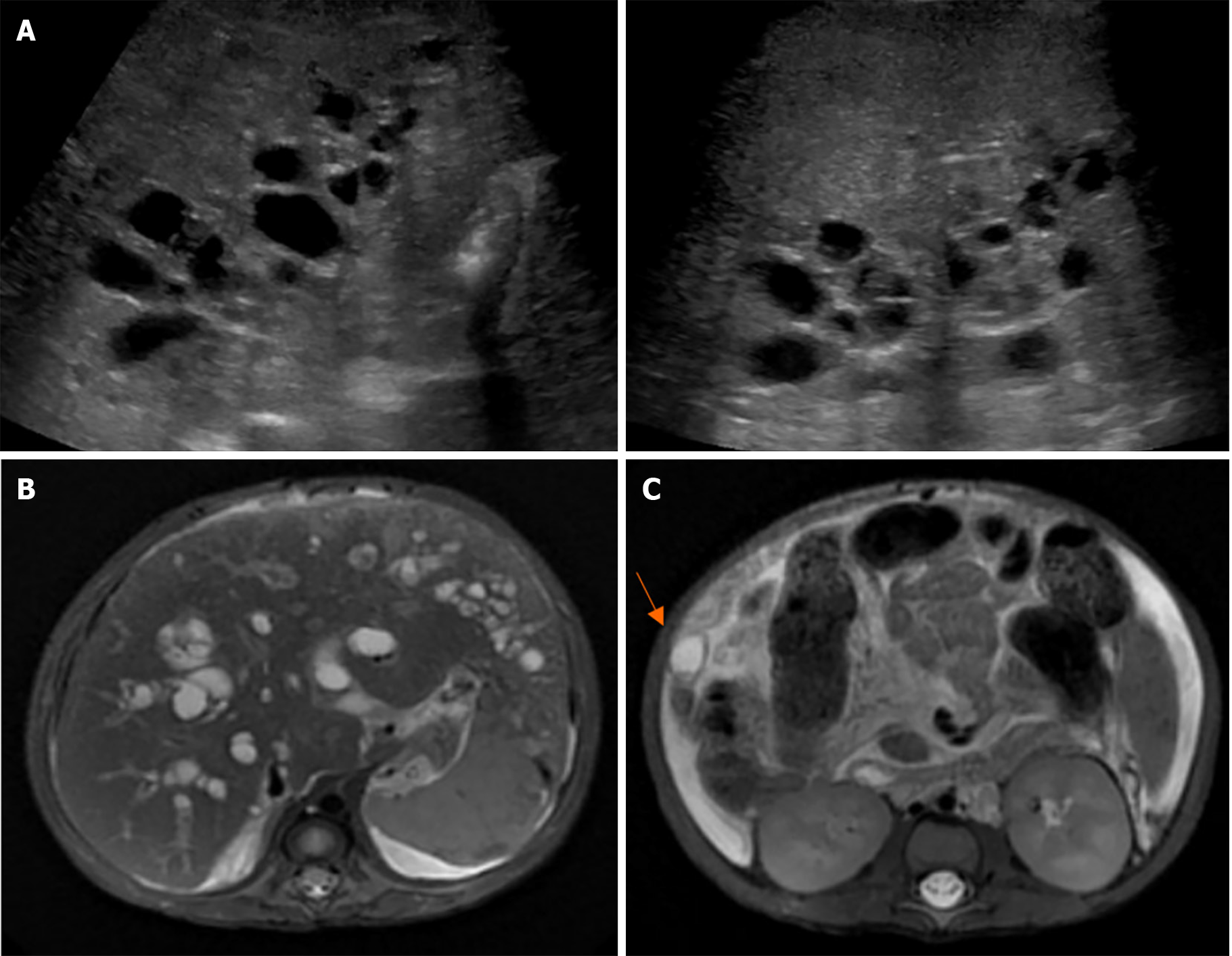
Figure 15 A 2-month-old male infant presented with jaundice and pale stools.
Exploratory laparotomy revealed a collapsed gallbladder, yellow bile, and patent right hepatic bile duct. The liver histopathology at the age of 2 months revealed cirrhosis with cholestasis jaundice. At the age of 7 months, repeated ultrasonography showed liver cirrhosis with evidence of portal hypertension, multiple cysts along the periportal distribution with a hyperechogenic area at the periportal region, probably a periportal type of Caroli disease, enlarged and diffusely increased parenchymal echogenicity of both kidneys, splenomegaly, and a moderate amount of ascites. The MRCP findings were diffuse cystic dilatation of bilateral intrahepatic bile ducts with the presence of a central dot sign at the peripheral zone and evidence of liver cirrhosis with portal hypertension. The patient underwent liver transplantation with a favorable outcome and a follow-up period of 10 years. A: Ultrasonography shows liver cirrhosis with multiple cysts along the periportal distribution, probably a periportal type of Caroli disease; B: Magnetic resonance cholangiopancreatography shows diffuse cystic dilatation of bilateral intrahepatic bile ducts; C: A 1.1 cm probable hepatic cyst at segment VI with a central dot sign.
- Citation: Eiamkulbutr S, Tubjareon C, Sanpavat A, Phewplung T, Srisan N, Sintusek P. Diseases of bile duct in children. World J Gastroenterol 2024; 30(9): 1043-1072
- URL: https://www.wjgnet.com/1007-9327/full/v30/i9/1043.htm
- DOI: https://dx.doi.org/10.3748/wjg.v30.i9.1043


