Copyright
©The Author(s) 2024.
World J Gastroenterol. Mar 7, 2024; 30(9): 1043-1072
Published online Mar 7, 2024. doi: 10.3748/wjg.v30.i9.1043
Published online Mar 7, 2024. doi: 10.3748/wjg.v30.i9.1043
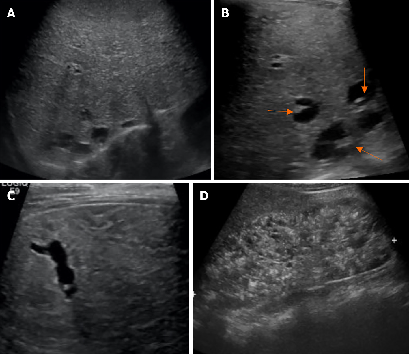
Figure 14 A 32-wk-old female infant with a family history of consanguinity, presented with an abdominal mass.
She was diagnosed with Calori syndrome (congenital hepatic fibrosis, choledochal cyst type V, and multiple cysts in the kidneys). Abdominal ultrasonography revealed enlargement of the left hepatic lobe, coarse parenchymal echotexture with a periportal hyperechoic band, small cysts in a posterosuperior segment of the right hepatic lobe, and marked enlargement of both kidneys with evidence suggestive of multi-cystic kidney disease. She had upper gastrointestinal bleeding from esophageal varices that can be controlled with esophageal ligation at the 8-year follow-up. A: Diffusely enlarged liver and coarse parenchymal echogenicity with several round and tubular cystic-like structures; B: Focal intrahepatic dilatation with central dot sign; C: Tubular cystic-like structures; D: Markedly enlarged kidneys with loss of corticomedullary differentiation, and multiple cysts in the medulla and cortices.
- Citation: Eiamkulbutr S, Tubjareon C, Sanpavat A, Phewplung T, Srisan N, Sintusek P. Diseases of bile duct in children. World J Gastroenterol 2024; 30(9): 1043-1072
- URL: https://www.wjgnet.com/1007-9327/full/v30/i9/1043.htm
- DOI: https://dx.doi.org/10.3748/wjg.v30.i9.1043


