Copyright
©The Author(s) 2024.
World J Gastroenterol. Mar 7, 2024; 30(9): 1043-1072
Published online Mar 7, 2024. doi: 10.3748/wjg.v30.i9.1043
Published online Mar 7, 2024. doi: 10.3748/wjg.v30.i9.1043
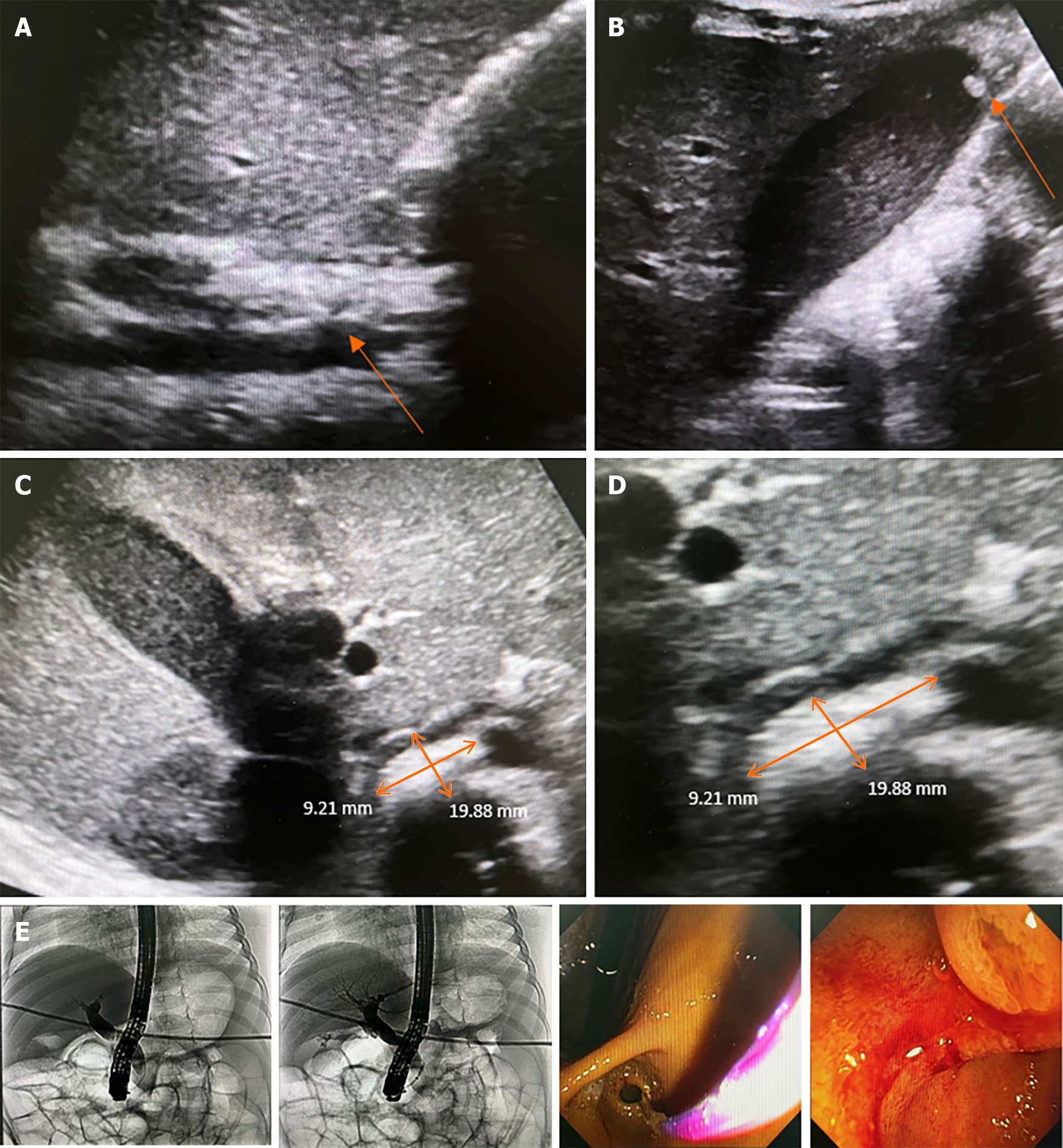
Figure 7 A 2-month-old female infant with anemia caused by chronic hemolysis.
Genetic analysis demonstrated heterozygous PKLR1 mutation/large deletion that was compatible with pyruvate kinase deficiency. Subsequently, she received regular blood transfusions every 3-4 wk. At the age of 3 years, the patient experienced intermittent abdominal pain, nausea, jaundice, and decreased appetite with no fever. Liver function test showed total bilirubin (TB) of 33.4, direct bilirubin (DB) of 31.2 mg/dL, aspartate aminotransferase of 270, alanine transaminase of 530, and alkaline phosphatase of 277 U/L. Abdominal ultrasonography showed marked splenomegaly, a dilated common bile duct (CBD) up to 0.9 cm in diameter, mild wall thickening, a suspected 2 cm × 0.9 cm distal CBD stone - well-distended gallbladder with bile sludge, and a 0.4 cm gallstone in the fundus. Subsequently, endoscopic retrograde cholangiography (ERCP) was performed, which revealed a bulging ampulla and CBD dilatation of 1 cm. The CRE balloon (12-15 mm) filled with 40 mL of air was placed for 60 s for sphincteroplasty. Later, balloon sweeping was performed, and small, blackish-pigmented stones were observed. After stone removal, the liver function test improved gradually as TB/DB 7.84/4.57 at 1 wk and normal values at 1 month. A: Dilated CBD, up to 9 mm, with mild wall thickening; B: A well-distended gallbladder with bile sludge and 0.4 cm stone at the fundus; C: A 2 cm × 0.9 cm distal CBD stone; D: A 2 cm × 0.9 cm distal CBD stone (zoom in); E: ERCP with CBD dilation with 12-15 mm CRE balloon filled with 40 mL of air placed for 60 s as sphincteroplasty.
- Citation: Eiamkulbutr S, Tubjareon C, Sanpavat A, Phewplung T, Srisan N, Sintusek P. Diseases of bile duct in children. World J Gastroenterol 2024; 30(9): 1043-1072
- URL: https://www.wjgnet.com/1007-9327/full/v30/i9/1043.htm
- DOI: https://dx.doi.org/10.3748/wjg.v30.i9.1043


