Copyright
©The Author(s) 2024.
World J Gastroenterol. Mar 7, 2024; 30(9): 1043-1072
Published online Mar 7, 2024. doi: 10.3748/wjg.v30.i9.1043
Published online Mar 7, 2024. doi: 10.3748/wjg.v30.i9.1043
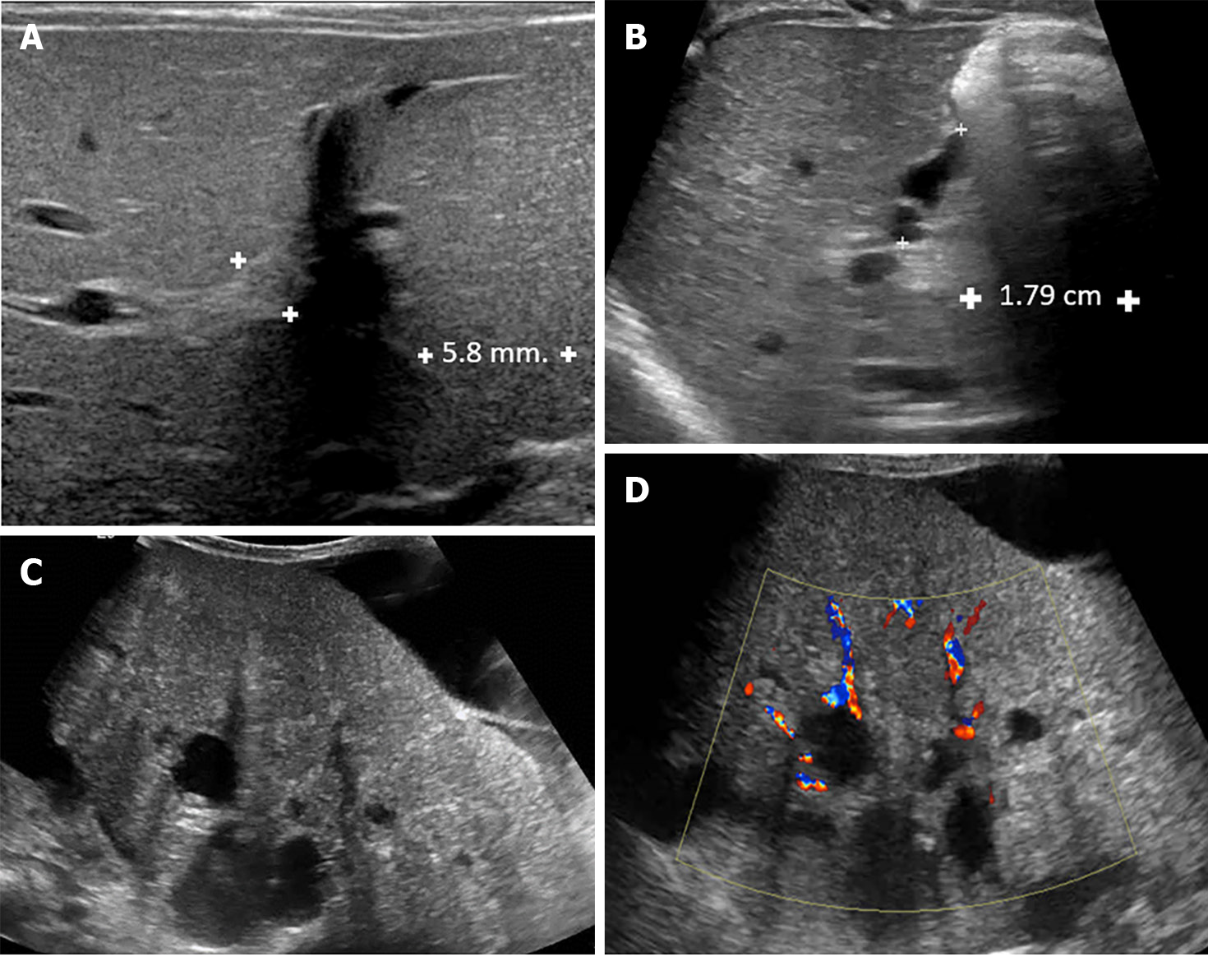
Figure 3 Ultrasound finding of biliary atresia.
A: A 25-d-old boy presented with prolong jaundice. Ultrasonography of the upper abdomen showed a thick echogenic band located anterior to the right portal vein and measuring approximately 5.8 mm in thickness, representing a positive triangular cord sign; B: A 49-d-old boy presented with prolonged jaundice and pale stools. Ultrasonography of the upper abdomen revealed: (1) A small size of gallbladder, measuring a maximal length of approximately 15.4 mm; (2) Lack of smooth, complete echogenic mucosal lining with an indistinct wall; and (3) Irregular gallbladder contour. These findings are consistent with a positive gallbladder ghost triad. The common bile duct was not visualized; C and D: An 11-month-old boy presented with clinical obstructive jaundice with abdominal distension. Ultrasonography of the upper abdomen revealed multiple irregular cystic lesions of various sizes in the central region of the right and left hepatic lobes and at the porta hepatis. Some cyst had echogenic content. Liver cirrhosis and ascites were noted.
- Citation: Eiamkulbutr S, Tubjareon C, Sanpavat A, Phewplung T, Srisan N, Sintusek P. Diseases of bile duct in children. World J Gastroenterol 2024; 30(9): 1043-1072
- URL: https://www.wjgnet.com/1007-9327/full/v30/i9/1043.htm
- DOI: https://dx.doi.org/10.3748/wjg.v30.i9.1043


